Microscopia a scansione di sonda

La microscopia a scansione di sonda (SPM, Scanning Probe Microscopy) è un ramo della microscopia che forma le immagini di superfici usando una sonda fisica che esegue la scansione del campione. Un'immagine della superficie è ottenuta meccanicamente spostando la sonda in un raster di scansione (raster scan) del campione, riga per riga e registrando l'interazione sonda-superficie in funzione della posizione. La SPM è stata istituita nel 1981 con l'invenzione del microscopio a scansione a effetto tunnel.

Molti microscopi a scansione di sonda sono in grado di raffigurare molte interazioni simultaneamente. La maniera di utilizzare queste interazioni per ottenere un'immagine è generalmente chiamata "modo".

La risoluzione varia alquanto da tecnica a tecnica, ma alcune tecniche di sonda raggiungono una risoluzione atomica piuttosto impressionante. Ciò è dovuto in gran parte alla capacità degli attuatori piezoelettrici di eseguire movimenti con una precisione e accuratezza a livello atomico o meglio in base al comando elettronico. Si potrebbe giustamente chiamare questa tipo di tecniche "piezoelettriche". L'altro denominatore comune è che i dati sono in genere ottenuti come una griglia bidimensionale di punti dati (data points), visualizzati in colori falsi come immagine del computer.

I tipi di microscopia a scansione di sonda

Di queste tecniche l'AFM e la STM sono quelle più comunemente utilizzate per misurazioni grossolane.

Punte della sonda

Le punte della sonda sono normalmente in platino/iridio oppure oro. Ci sono due metodi principali per ottenere una punta acuminata di sonda: l'incisione tramite acido e il taglio. Il primo consiste nell'immergere l'estremità di un filo prima in un bagno acido e aspettare fino a quando non viene inciso e la parte inferiore non sia sgocciolata del tutto. Il rimanente viene poi rimosso e la punta che ne risulta è spesso del diametro di un atomo. Un metodo alternativo e molto più rapido è quello di prendere un filo sottile e tagliarlo con un paio di forbici o un bisturi. Testare la punta prodotta tramite questo metodo su un campione con un profilo noto indicherà se la punta è buona o meno e un unico punto acuminato viene raggiunto in circa il 50% del tempo. Non è raro che tramite questo metodo risulti una punta con più di un picco; si può facilmente discernere che questa scansione causi un elevato livello di immagini fantasma.

Vantaggi della microscopia a scansione di sonda

  • La risoluzione dei microscopi non è limitata dalla diffrazione, ma solo dalle dimensioni del volume di interazione sonda-campione (vale a dire la funzione di diffusione di un punto), che può essere piccolo di alcuni picometri. Perciò è incomparabile la capacità di misurare le piccole differenze locali riguardo all'altezza dell'oggetto (come quella dei "gradini" (steps) di 135 picometri su <100> di silicio). Lateralmente l'interazione sonda-campione si estende solo attraverso l'atomo di punta o gli atomi coinvolti nell'interazione.
  • L'interazione può essere usata per modificare il campione onde poter creare strutture di piccole dimensioni (nanolitografia).
  • A differenza dei metodi utilizzati per il microscopio elettronico, i campioni non necessitano di un vuoto parziale, ma possono essere osservati in aria, a temperatura e pressione standard oppure mentre sono immersi in un recipiente per la reazione di liquidi.

Svantaggi della microscopia a scansione di sonda

  • La forma dettagliata della punta di scansione è a volte difficile da determinare. Il suo effetto sui dati che ne risultano è particolarmente rilevante se il campione varia notevolmente in altezza su distanze laterali di 10 nm o anche meno.
  • Le tecniche di scansione sono generalmente più lente ad acquisire le immagini, a causa del processo di scansione. Come risultato, gli sforzi vengono fatti per migliorare notevolmente la velocità di scansione. Come tutte le tecniche di scansione, l'inserimento di informazioni spaziali in una sequenza temporale apre la porta alle incertezze in metrologia, vale a dire di distanze e angoli laterali, che sorgono a causa degli effetti del dominio temporale come l'accumulo del campione, l'oscillazione del loop di retroazione (feedback) e le vibrazioni meccaniche.
  • La dimensione massima dell'immagine è generalmente più piccola.
  • La microscopia a scansione di sonda spesso non è utile per esaminare le immersioni di interfacce solido-solido o liquido-liquido.

Note

  1. ^ (EN) G. Binnig, C. F. Quate e Ch. Gerber, Atomic Force Microscope, in Physical Review Letters, vol. 56, n. 9, 3 marzo 1986, p. 930, DOI:10.1103/PhysRevLett.56.930.
  2. ^ (EN) W. J. Kaiser e L. D. Bell, Direct investigation of subsurface interface electronic structure by ballistic-electron-emission microscopy, in Physical Review Letters, vol. 60, n. 14, 1988, p. 1406, DOI:10.1103/PhysRevLett.60.1406.
  3. ^ (EN) L. Zhang, T. Sakai, N. Sakuma, T. Ono, K. Nakayama, Nanostructural conductivity and surface-potential study of low-field-emission carbon films with conductive scanning probe microscopy, in Applied Physics Letters, vol. 75, n. 22, 1999, pp. 3527-3529, DOI:10.1063/1.125377.
  4. ^ (EN) J. M. R. Weaver, David W. Abraham, High resolution atomic force microscopy potentiometry, in Journal of Vacuum Science & Technology B: Microelectronics and Nanometer Structures, vol. 9, n. 3, 1991, pp. 1559-1561, DOI:10.1116/1.585423.
  5. ^ (EN) S. R. Higgins, R. J. Hamers, Morphology and dissolution processes of metal sulfide minerals observed with the electrochemical scanning tunneling microscope, Madison (Wisconsin), marzo 1996, vol. 14, AVS, marzo 1996, pp. 1360-1364, DOI:10.1116/1.589098. URL consultato il 28 marzo 2010 (archiviato dall'url originale il 5 marzo 2016).
  6. ^ (EN) M. Fritz, M. Radmacher, N. Petersen, H. E. Gaub, Visualization and identification of intracellular structures by force modulation microscopy and drug induced degradation, in The 1993 international conference on scanning tunneling microscopy, The 1993 international conference on scanning tunneling microscopy, vol. 12, Beijing, China, AVS, maggio 1994, pp. 1526-1529, DOI:10.1116/1.587278. URL consultato il 28 marzo 2010 (archiviato dall'url originale il 5 marzo 2016).
  7. ^ (EN) M. Nonnenmacher, M. P. O'Boyle, H. K. Wickramasinghe, Kelvin probe force microscopy, in Applied Physics Letters, vol. 58, n. 25, 1991, pp. 2921-2923, DOI:10.1063/1.105227.
  8. ^ (EN) U. Hartmann, Magnetic force microscopy: Some remarks from the micromagnetic point of view, in Journal of Applied Physics, vol. 64, n. 3, 1988, pp. 1561-1564, DOI:10.1063/1.341836.
  9. ^ (EN) J. A. Sidles, J. L. Garbini, K. J. Bruland, D. Rugar, O. Züger, S. Hoen, C. S. Yannoni, Magnetic resonance force microscopy, in Reviews of Modern Physics, vol. 67, n. 1, 1995, p. 249, DOI:10.1103/RevModPhys.67.249.
  10. ^ (EN) E. BETZIG, J. K. TRAUTMAN, T. D. HARRIS, J. S. WEINER, R. L. KOSTELAK, Breaking the Diffraction Barrier: Optical Microscopy on a Nanometric Scale, in Science, vol. 251, n. 5000, 22 marzo 1991, pp. 1468-1470, DOI:10.1126/science.251.5000.1468. URL consultato il 5 ottobre 2009.
  11. ^ (EN) A. Roelofs, U. Bottger, R. Waser, F. Schlaphof, S. Trogisch, L. M. Eng, Differentiating 180° and 90° switching of ferroelectric domains with three-dimensional piezoresponse force microscopy, in Applied Physics Letters, vol. 77, n. 21, 2000, pp. 3444-3446, DOI:10.1063/1.1328049.
  12. ^ (EN) R. C. Reddick, R. J. Warmack, T. L. Ferrell, New form of scanning optical microscopy, in Physical Review B, vol. 39, n. 1, 1º gennaio 1989, p. 767, DOI:10.1103/PhysRevB.39.767. URL consultato il 5 ottobre 2009.
  13. ^ (EN) J. R. Matey, J. Blanc, Scanning capacitance microscopy, in Journal of Applied Physics, vol. 57, n. 5, 1985, pp. 1437-1444, DOI:10.1063/1.334506.
  14. ^ (EN) M. A. Eriksson, R. G. Beck, M. Topinka, J. A. Katine, R. M. Westervelt, K. L. Campman, A. C. Gossard, Cryogenic scanning probe characterization of semiconductor nanostructures, in Applied Physics Letters, vol. 69, n. 5, 29 luglio 1996, pp. 671-673, DOI:10.1063/1.117801. URL consultato il 5 ottobre 2009 (archiviato dall'url originale il 5 marzo 2016).
  15. ^ (EN) PK Hansma, B Drake, O Marti, SA Gould, CB Prater, The scanning ion-conductance microscope, in Science, vol. 243, n. 4891, 3 febbraio 1989, pp. 641-643, DOI:10.1126/science.2464851. URL consultato il 5 ottobre 2009.
  16. ^ (EN) R. Wiesendanger, M. Bode, Nano- and atomic-scale magnetism studied by spin-polarized scanning tunneling microscopy and spectroscopy, in Solid State Communications, vol. 119, n. 4-5, 25 luglio 2001, pp. 341-355, DOI:10.1016/S0038-1098(01)00103-X, ISSN 0038-1098 (WC · ACNP). URL consultato il 5 ottobre 2009.
  17. ^ (EN) P. De Wolf, J. Snauwaert, T. Clarysse, W. Vandervorst, L. Hellemans, Characterization of a point-contact on silicon using force microscopy-supported resistance measurements, in Applied Physics Letters, vol. 66, n. 12, 1995, pp. 1530-1532, DOI:10.1063/1.113636.
  18. ^ (EN) J. B. Xu, K. Lauger, K. Dransfeld, I. H. Wilson, Thermal sensors for investigation of heat transfer in scanning probe microscopy, in Review of Scientific Instruments, vol. 65, n. 7, 1994, pp. 2262-2266, DOI:10.1063/1.1145225.
  19. ^ (EN) G. Binnig, H. Rohrer, Ch. Gerber, E. Weibel, Tunneling through a controllable vacuum gap, in Applied Physics Letters, vol. 40, n. 2, 1982, pp. 178-180, DOI:10.1063/1.92999.
  20. ^ (EN) T. Trenkler, P. De Wolf, W. Vandervorst, L. Hellemans, Nanopotentiometry: Local potential measurements in complementary metal--oxide--semiconductor transistors using atomic force microscopy, in J. Vac. Sci. Techn. B, vol. 16, 1998, pp. 367-372, DOI:10.1116/1.589812.
  21. ^ (EN) A. M. Chang, H. D. Hallen, L. Harriott, H. F. Hess, H. L. Kao, J. Kwo, R. E. Miller, R. Wolfe, J. van der Ziel, T. Y. Chang, Scanning Hall probe microscopy, in Applied Physics Letters, vol. 61, n. 16, 1992, pp. 1974-1976, DOI:10.1063/1.108334.

Altri progetti

Collegamenti esterni

Controllo di autoritàGND (DE4330328-6 · NDL (ENJA00577272

Read other articles:

British television series Fool Me OnceGenreThrillerBased onFool Me Onceby Harlan CobenWritten by Danny Brocklehurst Charlotte Coben Yemi Oyefuwa Nina Metivier Tom Farrelly Directed byDavid MooreNimer RashedStarring Michelle Keegan Dino Fetscher Richard Armitage Joanna Lumley Adeel Akhtar Emmett J. Scanlan Marcus Garvey Dänya Griver Natalie Anderson Daniel Burt Adelle Leonce Joe Armstrong Theme music composerDavid BuckleyLuke Richards[1]Country of originUnited KingdomOriginal language...


Roman historian and theologian (c. 375/385 – c. 420 AD) For other uses, see Orosius (disambiguation). OrosiusMiniature from the Saint-Epure codexBornc. 375/85 ADBraga, GallaeciaDiedc. 420 ADOccupation(s)Theologian and historianAcademic backgroundInfluences Augustine of Hippo Livy Jerome Junianus Justinus Tacitus Suetonius Florus Academic workMain interests Providentialism Universal history Germanic paganism Paulus Orosius (/ˈpɔːləs əˈroʊʒəs/; born c. 375/385 – c. 420 AD),[1&#...
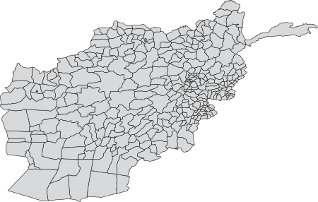
 

Artikel ini membutuhkan rujukan tambahan agar kualitasnya dapat dipastikan. Mohon bantu kami mengembangkan artikel ini dengan cara menambahkan rujukan ke sumber tepercaya. Pernyataan tak bersumber bisa saja dipertentangkan dan dihapus.Cari sumber: Distrik di Afganistan – berita · surat kabar · buku · cendekiawan · JSTOR (Januari 2019) Afganistan Artikel ini adalah bagian dari seri Politik dan KetatanegaraanAfganistan Eksekutif Amir (daftar) Hibatullah ...

  لمعانٍ أخرى، طالع بيرو (توضيح).   جمهورية بيرو República del Perú  (إسبانية) بيروعلم بيرو بيروشعار بيرو الشعار الوطني(بالإسبانية: Firme y feliz por la unión)‏  النشيد: نشيد بيرو الوطني الأرض والسكان إحداثيات 9°24′S 76°00′W / 9.4°S 76°W / -9.4; -76   [1] أعلى قمة هوسكاران...


German-Iranian actress Some of this article's listed sources may not be reliable. Please help improve this article by looking for better, more reliable sources. Unreliable citations may be challenged and removed. (June 2023) (Learn how and when to remove this template message) Narges RashidiRashidi at the U.S. Embassy Berlin during salute to the Berlinale 15.2.2016BornNarges Rashidi (1980-03-21) 21 March 1980 (age 44)Khorramabad, IranOccupationActressYears active2004–presentHeight...


Ethnic group For other uses, see Asháninka (disambiguation). This article needs additional citations for verification. Please help improve this article by adding citations to reliable sources. Unsourced material may be challenged and removed.Find sources: Asháninka – news · newspapers · books · scholar · JSTOR (August 2017) (Learn how and when to remove this template message) AsháninkaAshenikaTotal population99,122 (2014)Regions with significant pop...

United States Navy admiral This article needs additional citations for verification. Please help improve this article by adding citations to reliable sources. Unsourced material may be challenged and removed.Find sources: Clark H. Woodward – news · newspapers · books · scholar · JSTOR (June 2020) (Learn how and when to remove this template message) Clark H. WoodwardRADM Clark H. Woodward in 1919Birth nameClark Howell WoodwardNickname(s)WoodyBorn(1877-0...


Exploitable weakness in a computer system Part of a series onComputer hacking History Phreaking Cryptovirology Hacking of consumer electronics List of hackers Hacker culture and ethic Hackathon Hacker Manifesto Hackerspace Hacktivism Maker culture Types of hackers Black hat Grey hat White hat Conferences Black Hat Briefings Chaos Communication Congress DEF CON Hackers on Planet Earth Security BSides ShmooCon Summercon Computer crime Crimeware List of computer criminals Script kiddie Hacking t...


American psychiatrist and retired military officer Loree SuttonCommissioner of the New York City Department of Veterans' ServicesIn officeJuly 2016 – November 2019Appointed byBill de BlasioSucceeded byJames Hendon Personal detailsBorn (1959-07-15) July 15, 1959 (age 64)Loma Linda, California, U.S.Political partyDemocraticSpouse Laurie Leitch ​(m. 2015)​[1]EducationPacific Union College (BS)Loma Linda University (MD)National War College (MS)...

1993 film by R. V. Udayakumar YajamanTheatrical release posterDirected byR. V. UdayakumarScreenplay byR. V. UdayakumarStory bySujatha UdayakumarProduced byM. SaravananM. BalasubramaniamM. S. GuhanStarringRajinikanthMeenaAishwaryaCinematographyKarthik RajaEdited byB. S. NagarajMusic byIlaiyaraajaProductioncompanyAVM ProductionsRelease date 18 February 1993 (1993-02-18) Running time153 minutesCountryIndiaLanguageTamil Yajaman (transl. Figure Head) is a 1993 Indian Tamil-lan...


2022年內華達州副州長選舉 ← 2018 2022年11月8日 2026 →   获提名人 斯塔夫羅斯·安東尼 麗莎·卡諾·伯克黑德 政党 共和黨 民主黨 民選得票 500994 463871 得票率 49.4% 45.8% 縣市結果安東尼:     40–50%     50–60%     60–70%     70–80%     80–90%伯克黑德:     40–50% 选前副州長 麗莎...


Depiction of Ancamna and Mars Smertrius from Freckenfeld in the ancient territory of the Nemetes. In Gallo-Roman religion, Ancamna was a goddess worshipped particularly in the valley of the river Moselle. She was commemorated at Trier and Ripsdorf as the consort of Lenus Mars,[1] and at Möhn as the consort of Mars Smertulitanus.[2][3] At Trier, altars were set up in honour of Lenus Mars, Ancamna and the genii of various pagi of the Treveri, giving the impression of Le...

2020年夏季奥林匹克运动会波兰代表團波兰国旗IOC編碼POLNOC波蘭奧林匹克委員會網站olimpijski.pl(英文)(波兰文)2020年夏季奥林匹克运动会(東京)2021年7月23日至8月8日(受2019冠状病毒病疫情影响推迟,但仍保留原定名称)運動員206參賽項目24个大项旗手开幕式:帕维尔·科热尼奥夫斯基(游泳)和马娅·沃什乔夫斯卡(自行车)[1]闭幕式:卡罗利娜·纳亚(皮划艇)&#...
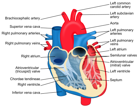
 

Untuk kegunaan lain dari bilik, lihat Bilik. Bilik jantung yang merupakan ruang terbesar dari jantung. Bilik jantung atau ventrikel adalah ruang jantung yang mempunyai tanggung jawab untuk memompa darah meninggalkan jantung[1] Sumber lain menjelaskan bahwa bilik jantung adalah dua bilik besar yang tugasnya menerima darah dari serambi jantung dan juga berkontraksi untuk memompa darah yang berada di dalam keluar jantung dan ke seluruh organ tubuh.[2] Pada mamalia (dalam kategori...


Area of tissue in the eye This article needs additional citations for verification. Please help improve this article by adding citations to reliable sources. Unsourced material may be challenged and removed.Find sources: Trabecular meshwork – news · newspapers · books · scholar · JSTOR (December 2008) (Learn how and when to remove this message) Trabecular meshworkEnlarged general view of the iridial angle. (When enlarged, visible with older label of 't...
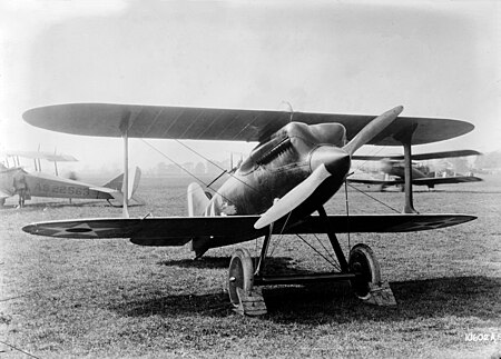
Model 23, CR, R-6 The CR-1 with Bert Acosta, 1921 Role Racing aircraftType of aircraft Manufacturer Curtiss Aeroplane and Motor Company First flight 1 August 1921 Primary user United States Navy Number built 4 The Curtiss CR was a racing aircraft designed for the United States Navy in 1921 by Curtiss. It was a conventional single-seater biplane with a monocoque fuselage and staggered single-bay wings of equal span braced with N-struts. Two essentially similar landplane versions were bui...


US 2-cent stamp of 1870, cancelled with a leaf shape in blue ink A fancy cancel is a postal cancellation that includes an artistic design. Although the term may be used of modern machine cancellations that include artwork, it primarily refers to the designs carved in cork and used in 19th century post offices of the United States. When postage stamps were introduced in the US in 1847, postmasters were required to deface them to prevent reuse, but it was left up to them to decide exactly how t...


Frigate class of the Royal Navy Perseverance-class frigate Phoenix off Malta Class overview NamePerseverance-class frigate Operators Royal Navy Preceded byMinerva class Succeeded byPallas class Built1780–1783, 1801–1811 In service1781–1874 Planned12 Completed11 Cancelled1 Lost5 General characteristics first iteration[1] TypeFifth-rate frigate Tons burthen871 42⁄94 (bm) Length 137 ft (41.8 m) (gundeck) 113 ft 5+1⁄2 in (34.6 m) (keel)...

Pterocaesio Pterocaesio pisang Klasifikasi ilmiah Kerajaan: Animalia Filum: Chordata Kelas: Actinopterygii Ordo: Perciformes Famili: caesionidae Genus: PterocaesioBleeker, 1876 Spesies Lihat teks Sinonim[1] Clupeolabrus Nichols, 1923 Liocaesio Bleeker, 1876 Pisinnicaesio Carpenter, 1987 Squamosicaesio Carpenter, 1987 Pterocaesio adalah genus ikan bersirip kipas laut dalam keluarga Caesionidae. Mereka berasal dari Samudra Hindia dan Samudra Pasifik bagian barat. Daftar spesies[2&#...


Unidad periférica de XánthiXánthi Unidad periférica Ubicación de la unidad periféricaCoordenadas 41°08′17″N 24°53′11″E / 41.1380289, 24.8862688Capital XánthiEntidad Unidad periférica • País Grecia • Periferia Macedonia Oriental y TraciaSuperficie   • Total 1793 km²Población (2001)   • Total 101 856 hab. • Densidad 60,34 hab./km²Huso horario UTC+02:00 y UTC+03:00Código postal 67x xxMatrícula AHI...