Cardiac cycle

Cardiac cycle
OrganismsAnimalia
Biological systemCirculatory system
HealthBeneficial
ActionInvoluntary
MethodBlood is allowed to enter relaxed ventricle chamber from vein through venous valve. Heart muscle contracts ventricle and blood is expelled through arterial valve to artery.
OutcomeCirculation of blood per minute (Humans)
Duration0.6–1 second (Humans)
*Animalia with the exception of Porifera, Cnidaria, Ctenophora, Platyhelminthes, Bryozoan, Amphioxus.
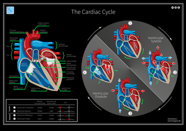
The cycle diagram depicts one heartbeat of the continuously repeating cardiac cycle, namely: ventricular diastole followed by ventricular systole, etc.—while coordinating with atrial systole followed by atrial diastole, etc. The cycle also correlates to key electrocardiogram tracings: the T wave (which indicates ventricular diastole); the P wave (atrial systole); and the QRS 'spikes' complex (ventricular systole)—all shown as color purple-in-black segments.[1][2]
The Cardiac Cycle: Valve Positions, Blood Flow, and ECG
The parts of a QRS complex and adjacent deflections. Re the cardiac cycle, atrial systole begins at the P wave; ventricular systole begins at the Q deflection of the QRS complex.

The cardiac cycle is the performance of the human heart from the beginning of one heartbeat to the beginning of the next.[1] It consists of two periods: one during which the heart muscle relaxes and refills with blood, called diastole, following a period of robust contraction and pumping of blood, called systole.[1] After emptying, the heart relaxes and expands to receive another influx of blood returning from the lungs and other systems of the body, before again contracting to pump blood to the lungs and those systems.

Assuming a healthy heart and a typical rate of 70 to 75 beats per minute, each cardiac cycle, or heartbeat, takes about 0.8 second to complete the cycle.[2][3] Duration of the cardiac cycle is inversely proportional to the heart rate.[4]

Description

There are two atrial and two ventricle chambers of the heart; they are paired as the left heart and the right heart—that is, the left atrium with the left ventricle, the right atrium with the right ventricle—and they work in concert to repeat the cardiac cycle continuously (see cycle diagram at right margin).[1] At the start of the cycle, during ventricular diastole–early, the heart relaxes and expands while receiving blood into both ventricles through both atria; then, near the end of ventricular diastole–late, the two atria begin to contract (atrial systole), and each atrium pumps blood into the ventricle below it.[5] During ventricular systole the ventricles contract and vigorously pulse (or eject) two separated blood supplies from the heart—one to the lungs and one to all other body organs and systems—while the two atria relax (atrial diastole). This precise coordination ensures that blood is efficiently collected and circulated throughout the body.[1][2]

The mitral and tricuspid valves, also known as the atrioventricular, or AV valves, open during ventricular diastole to permit filling. Late in the filling period the atria begin to contract (atrial systole) forcing a final crop of blood into the ventricles under pressure—see cycle diagram. Then, prompted by electrical signals from the sinoatrial node, the ventricles start contracting (ventricular systole), and as back-pressure against them increases the AV valves are forced to close, which stops the blood volumes in the ventricles from flowing in or out; this is known as the isovolumic contraction stage.[1][6]

Due to the contractions of the systole, pressures in the ventricles rise quickly, exceeding the pressures in the trunks of the aorta and the pulmonary arteries and causing the requisite valves (the aortic and pulmonary valves) to open—which results in separated blood volumes being ejected from the two ventricles.[1] This is the ejection stage of the cardiac cycle; it is depicted (see circular diagram) as the ventricular systole–first phase followed by the ventricular systole–second phase.[2] After ventricular pressures fall below their peak(s) and below those in the trunks of the aorta and pulmonary arteries, the aortic and pulmonary valves close again—see, at the right margin, Wiggers diagram, blue-line tracing.[2]

Next is the isovolumic relaxation, during which pressure within the ventricles begin to fall significantly, and thereafter the atria begin refilling as blood returns to flow into the right atrium (from the vena cavae) and into the left atrium (from the pulmonary veins).[1] As the ventricles begin to relax, the mitral and tricuspid valves open again, and the completed cycle returns to ventricular diastole and a new "Start" of the cardiac cycle.[1][2]

Throughout the cardiac cycle, blood pressure increases and decreases. The movements of cardiac muscle are coordinated by a series of electrical impulses produced by specialized pacemaker cells found within the sinoatrial node and the atrioventricular node. Cardiac muscle is composed of myocytes which initiate their internal contractions without receiving signals from external nerves—with the exception of changes in the heart rate due to metabolic demand.[1][2]

In an electrocardiogram, electrical systole initiates the atrial systole at the P wave deflection of a steady signal; and it starts contractions (systole).[2]

Cardiac cycle and Wiggers diagram

A Wiggers diagram illustrate events and details of the cardiac cycle with electrographic trace lines, which depict (vertical) changes in a parameter's value as time elapses left-to-right.[2] The ventricular "diastole", or relaxation, begins with "isovolumic relaxation", then proceeds through three sub-stages of inflow, namely: "rapid inflow", "diastasis", and "atrial systole". During the "diastole" period, the "ventricular volume" increases (see red-line tracing), beginning after the vertical bar at "Aortic valve closes" and ending with the vertical bar at R in the QRS complex. The ventricular "Systole", or contraction, begins with "isovolumic contraction", i.e., with the vertical bar at "A -V valve closes"; it ends with completing the "ejection" stage at the bar at "aortic valve closes". During "ejection" stage, the (red-line) tracing of "ventricular volume" falls to its least amount (see ejection fraction) as the ventricles pump blood to the pulmonary arteries and to the aorta.

The cardiac cycle involves four major stages of activity: 1) "isovolumic relaxation", 2) inflow, 3) "isovolumic contraction", 4) "ejection".[1][2] Stages 1 and 2 together—"isovolumic relaxation" plus inflow (equals "rapid inflow", "diastasis", and "atrial systole")—comprise the ventricular diastole period, including atrial systole, during which blood returning to the heart flows through the atria into the relaxed ventricles.[1][2] Stages 3 and 4 together—"isovolumic contraction" plus "ejection"—are the ventricular systole period, which is the simultaneous pumping of separate blood supplies from the two ventricles, one to the pulmonary artery and one to the aorta.[1][2] Notably, near the end of the diastole, the atria begin contracting, then pump blood into the ventricles; this pressurized delivery during ventricular relaxation (ventricular diastole) is called the atrial systole.[1][2]

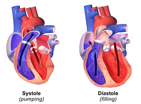
Diastole (at right) normally refers to atria and ventricles at relaxation and expansion together—while refilling with blood returning to the heart. Systole (left) typically refers to ventricular systole, during which the ventricles are pumping (or ejecting) blood out of the heart through the aorta and the pulmonary veins.

The closure of the aortic valve causes a rapid change in pressure in the aorta called the incisura. This short sharp change in pressure is rapidly attenuated down the arterial tree. The pulse wave form is also reflected from branches in the arterial tree and gives rise to a dicrotic notch in main arteries. The summation of the reflected pulse wave and the systolic wave may increase pulse pressure and help tissue perfusion. With increasing age, the aorta stiffens and can become less elastic which will reduce peak pulse in the periphery.

Physiology

CGI-animated graphic of the human heart, sectioned, with motions and timing synced with the Wiggers diagram. The section shows: 1) the opened ventricles contracting once per heartbeat—that is, once per each cardiac cycle; 2) the (partly obscured) mitral valve of the left heart; 3) the tricuspid and pulmonary valves of the right heart—note these paired valves open and close oppositely. + (The aortic valve of the left heart is located below the pulmonary valve, and is completely obscured.) The (unsectioned) atria are seen above the ventricles.

The heart is a four-chambered organ consisting of right and left halves, called the right heart and the left heart. The upper two chambers, the left and right atria, are entry points into the heart for blood-flow returning from the circulatory system, while the two lower chambers, the left and right ventricles, perform the contractions that eject the blood from the heart to flow through the circulatory system. Circulation is split into pulmonary circulation—during which the right ventricle pumps oxygen-depleted blood to the lungs through the pulmonary trunk and arteries; or the systemic circulation—in which the left ventricle pumps/ejects newly oxygenated blood throughout the body via the aorta and all other arteries.[1][2]

Heart electrical conduction system

In a healthy heart all activities and rests during each individual cardiac cycle, or heartbeat, are initiated and orchestrated by signals of the heart's electrical conduction system, which is the "wiring" of the heart that carries electrical impulses throughout the body of cardiomyocytes, the specialized muscle cells of the heart.[1][2] These impulses ultimately stimulate heart muscle to contract and thereby to eject blood from the ventricles into the arteries and the cardiac circulatory system; and they provide a system of intricately timed and persistent signaling that controls the rhythmic beating of the heart muscle cells, especially the complex impulse-generation and muscle contractions in the atrial chambers.

The rhythmic sequence (or sinus rhythm) of this signaling across the heart is coordinated by two groups of specialized cells, the sinoatrial (SA) node, which is situated in the upper wall of the right atrium, and the atrioventricular (AV) node located in the lower wall of the right heart between the atrium and ventricle.[1][2] The sinoatrial node, often known as the cardiac pacemaker, is the point of origin for producing a wave of electrical impulses that stimulates atrial contraction by creating an action potential across myocardium cells.[7][8]

Impulses of the wave are delayed upon reaching the AV node, which acts as a gate to slow and to coordinate the electrical current before it is conducted below the atria and through the circuits known as the bundle of His and the Purkinje fibers—all which stimulate contractions of both ventricles. The programmed delay at the AV node also provides time for blood volume to flow through the atria and fill the ventricular chambers—just before the return of the systole (contractions), ejecting the new blood volume and completing the cardiac cycle.[8] (See Wiggers diagram: "Ventricular volume" tracing (red), at "Systole" panel.)

Diastole and systole in the cardiac cycle

Cardiac diastole: Both AV valves (tricuspid in the right heart (light-blue), mitral in the left heart (pink)) are open to enable blood to flow directly into both left and right ventricles, where it is collected for the next contraction.
Cardiac (ventricular) systole: Both AV valves (tricuspid in the right heart (light-blue), mitral in the left heart (pink)) are closed by back-pressure as the ventricles are contracted and their blood volumes are ejected through the newly-opened pulmonary valve (dark-blue arrow) and aortic valve (dark-red arrow) into the pulmonary trunk and aorta respectively.

Cardiac diastole is the period of the cardiac cycle when, after contraction, the heart relaxes and expands while refilling with blood returning from the circulatory system.[1][2] Both atrioventricular (AV) valves open to facilitate the 'unpressurized' flow of blood directly through the atria into both ventricles, where it is collected for the next contraction. This period is best viewed at the middle of the Wiggers diagram—see the panel labeled "diastole". Here it shows pressure levels in both atria and ventricles as near-zero during most of the diastole. (See gray and light-blue tracings labeled "atrial pressure" and "ventricular pressure"—Wiggers diagram.) Here also may be seen the red-line tracing of "Ventricular volume", showing an increase in blood volume from the low plateau of the "isovolumic relaxation" stage to the maximum volume occurring in the "atrial systole" sub-stage.[1][2]

Atrial systole

Atrial systole is the contracting of cardiac muscle cells of both atria following electrical stimulation and conduction of electrical currents across the atrial chambers (see above, Physiology).[1][2] While nominally a component of the heart's sequence of systolic contraction and ejection, atrial systole actually performs the vital role of completing the diastole, which is to finalize the filling of both ventricles with blood while they are relaxed and expanded for that purpose. Atrial systole overlaps the end of the diastole, occurring in the sub-period known as ventricular diastole–late (see cycle diagram). At this point, the atrial systole applies contraction pressure to 'topping-off' the blood volumes sent to both ventricles; this atrial contraction closes the diastole immediately before the heart again begins contracting and ejecting blood from the ventricles (ventricular systole) to the aorta and arteries.[1][2]

Ventricular systole

Ventricular systole is the contractions, following electrical stimulations, of the ventricular syncytium of cardiac muscle cells in the left and right ventricles. Contractions in the right ventricle provide pulmonary circulation by pulsing oxygen-depleted blood through the pulmonary valve then through the pulmonary arteries to the lungs. Simultaneously, contractions of the left ventricular systole provide systemic circulation of oxygenated blood to all body systems by pumping blood through the aortic valve, the aorta, and all the arteries. (Blood pressure is routinely measured in the larger arteries off the left ventricle during the left ventricular systole).[1][2]

See also

References

  1. ^ a b c d e f g h i j k l m n o p q r s t u v Pollock JD, Makaryus AN (3 October 2022). "Physiology: Cardiac cycle". StatPearls Publishing, US National Library of Medicine. Retrieved 28 March 2024.
  2. ^ a b c d e f g h i j k l m n o p q r s t u Silverthorn, DU (10 November 2022). "Constructing the Wiggers diagram using core concepts: a classroom activity". Advances in Physiology Education. 46 (4). doi:10.1152/advan.00046.2022.
  3. ^ Gersh, Bernard J (2000). Mayo Clinic Heart Book. New York: William Morrow. pp. 6–8. ISBN 0-688-17642-9.
  4. ^ Hall JE, Hall ME (2011). Guyton and Hall Textbook of Medical Physiology (12th ed.). Philadelphia: Elsevier. pp. 104–105. ISBN 978-1-4160-4574-8.
  5. ^ Topol, Eric J (2000). Cleveland Clinic Heart Book. New York: Hyperion. pp. 4–5. ISBN 0-7868-6495-8.
  6. ^ Topol, Eric J (2000). Cleveland Clinic Heart Book. New York: Hyperion. pp. 8–10. ISBN 0-7868-6495-8.
  7. ^ Gersh, Bernard J (2000). Mayo Clinic Heart Book. New York: William Morrow. p. A12. ISBN 0-688-17642-9.
  8. ^ a b Bellenir, Karen (2000). Heart Diseases and Disorders Sourcebook, 2nd ed. Detroit: Frederick G. Ruffner, Jr./Omnigraphics. pp. 65–67. ISBN 0-7808-0238-1.

Bibliography

Read other articles:

У этого термина существуют и другие значения, см. Таловка. СелоТаловка 49°58′40″ с. ш. 45°02′07″ в. д.HGЯO Страна  Россия Субъект Федерации Волгоградская область Муниципальный район Камышинский Сельское поселение Таловское История и география Основан в 1812 году Час

سفين توفت معلومات شخصية الميلاد 9 مايو 1977 (العمر 46 سنة)كندا الطول 1.80 م (5 قدم 11 بوصة) الجنسية  كندا الوزن 74 كـغ (163 رطل؛ 11.7 ستون) الحياة العملية الدور دراج الفرق أوريكا سكوت (2012–2018)إي أف إديوكيشن نيبو (2009–2010)رالي سايكلينغ (2019–)  المهنة دراج  نوع السباق سبا...

Endive Escarole endive Klasifikasi ilmiah Kerajaan: Plantae Divisi: Magnoliophyta Kelas: Magnoliopsida Ordo: Asterales Famili: Asteraceae Genus: Cichorium Spesies: C. endivia Nama binomial Cichorium endiviaL. Belgian endive Belgian endive Endive (pengucapan: /ˈɛndaɪv/) atau andewi[1], Cichorium endivia adalah sayuran yang termasuk ke dalam familia Asteraceae. Endive dapat dimasak atau dimakan mentah dalam salad. Teknik menanam endive ditemukan secara tidak sengaja pada tahun 1...

Wu Kai Sha烏溪沙Stasiun angkutan cepat MTRNama TionghoaHanzi Tradisional 烏溪沙 Hanzi Sederhana 乌溪沙 Yale KantonWūkāisā Arti harfiahCrow brook sandTranskripsiTionghoa StandarHanyu PinyinWūxīshāYue: KantonRomanisasi YaleWūkāisāJyutpingWu1kai1saa1 Informasi umumLokasiBerdekatan dengan Lake Silver, Sai Sha Road, Ma On ShanDistrik Sha Tin, Hong KongKoordinat22°25′45″N 114°14′38″E / 22.4291°N 114.2438°E / 22.4291; 114.2438Koordinat: 22°25

ChivitoJenisRoti lapisTempat asalUruguayDibuat olehAntonio Carbonara[1]Bahan utamaRoti bulat, daging sapi churrasco, bakon, telur (goreng atau rebus), ham, zaitun hitam atau hijau, mozzarella, tomat, mayones  Media: Chivito Bahan baku pada roti lapis Chivito Sebuah roti lapis Chivito, dengan potongan seluruh bahan komposisinya Chivito adalah salah satu hidangan nasional dari Uruguay, yang terdiri dari potongan tipis steak daging sapi matang (churrasco), dengan mozzarella, tom...

Tom Brokaw Brokaw in 2007 Achtergrondinformatie Geboorteplaats Webster (South Dakota) Beroep Journalist, schrijver (en) IMDb-profiel Portaal    Media Tom Brokaw, geboren als Thomas John Brokaw (Webster (South Dakota), 6 februari 1940), is een Amerikaans journalist, tv-presentator en schrijver. Hij werd vooral bekend als anchorman van de nieuwsrubriek Nightly News van het televisienetwerk NBC. Daarnaast kwam hij ook met andere tv-producties en schreef hij boeken en bijdragen voor kra...

FleabagJudul FleabagGenre Drama komedi Tragikomedi PembuatPhoebe Waller-BridgeDitulis olehPhoebe Waller-BridgeSutradara Harry Bradbeer Tim Kirkby (e. 1) Pemeran Phoebe Waller-Bridge Sian Clifford Olivia Colman Bill Paterson Brett Gelman Hugh Skinner Hugh Dennis Ben Aldridge Jamie Demetriou Jenny Rainsford Andrew Scott Fiona Shaw Kristin Scott Thomas Ray Fearon Penata musikIsobel Waller-BridgeNegara asalBritania RayaBahasa asliInggrisJmlh. seri2Jmlh. episode12 (daftar episode)ProduksiPro...

Antonov A-40 Krylya Tanka (Rusia: крылья танка, yang berarti sayap tank) adalah upaya Soviet untuk memungkinkan tank untuk meluncur ke medan perang setelah ditarik tinggi-tinggi oleh pesawat terbang, untuk mendukung pasukan udara atau partisan. Sebuah prototipe dibangun dan diuji pada tahun 1942, tetapi tidak dapat diterapkan. Kendaraan ini kadang-kadang disebut A-40T atau KT. Spesifikasi Karakteristik umum Kru: Dua Kapasitas: 1 × T-60 tank Panjang: 12.06 m (39 ft 6 ¾ in) Le...

Coordenadas: 46° 25' N 10° 21' E Valdisotto    Comuna   Localização ValdisottoLocalização de Valdisotto na Itália Coordenadas 46° 25' N 10° 21' E Região Lombardia Província Sondrio Características geográficas Área total 88 km² População total 3 216 hab. Densidade 37 hab./km² Altitude 1 120 m Outros dados Comunas limítrofes Bormio, Grosio, Sondalo, Valdidentro, Valfurva Código ISTAT 014072 Código cadastral L563 C...

2018 International Wrestling Revolution Group event Rebelión de los Juniors (2015)Official poster showing all eight main event competitorsPromotionInternational Wrestling Revolution GroupDateMarch 1, 2015[1]CityNaucalpan, State of Mexico[1]VenueArena Naucalpan[1]Event chronology ← PreviousEl Protector Next →Guerra del Golfo Rebelión de los Juniors chronology ← Previous2014 Next →2016 Rebelión de los Juniors (2015) (Spanish for The Junior ...

У Вікіпедії є статті про інші значення цього терміна: 4-та армія. 4-та армія (Велика Британія)Fourth Army (United Kingdom) Емблема 4-ї британської арміїНа службі 5 лютого 1916 — початок 1919Країна  Велика БританіяВид Британська арміяРоль Сухопутні військаЧисельність загальновійськов...

County in Alabama, United States County in AlabamaTuscaloosa CountyCountyTuscaloosa County Courthouse in TuscaloosaLocation within the U.S. state of AlabamaAlabama's location within the U.S.Coordinates: 33°12′23″N 87°32′05″W / 33.2065°N 87.5346°W / 33.2065; -87.5346Country United StatesState AlabamaFoundedFebruary 6, 1818[1]Named forTuskaloosaSeatTuscaloosaLargest cityTuscaloosaArea • Total1,351 sq mi (3,500 km2)...

Detail of an infant's bodice in Limerick lace Needlerun net is a family of laces created by using a needle to embroider on a net ground.[1] Along with Tambour lace this became more popular with the advent of machine made netting. It is used in Limerick lace. References ^ Pat Earnshaw. A Dictionary of Lace. Shire Publications. ISBN 0-85263-700-4. vteLace typesNeedle lace Filet lace Punto in Aria Point de Venise Point de France Alençon Argentan Argentella Armenian Halas lace Hedeb...

Sede central de CAPES em Brasília El CAPES (en portugués, Coordenação de aperfeiçoamento de pessoal de nivel superior) es un organismo brasileño bajo la autoridad del Ministerio de Educación que desempeña tres actividades principales: la evaluación de los programas brasileños de postgrado, el pago de becas y auxilios a investigadores y sobre todo a estudiantes de maestría y doctorado, y el mantenimiento de un Portal de Periódicos que incluye más de 12.000 títulos, la mayor parte...

Modification of Android devices to gain rooted access Not to be confused with bootloader unlocking or SIM unlocking.Rooting is the process by which users of Android devices can attain privileged control (known as root access) over various subsystems of the device, usually smartphones. Because Android is based on a modified version of the Linux kernel, rooting an Android device gives similar access to administrative (superuser) permissions as on Linux or any other Unix-like operating system su...

Planned LEO station designed by Nanoracks Starlab is a planned LEO (low Earth orbit) space station designed by Nanoracks for commercial space activities uses, whose launch is planned for 2028. History Background In March 2021, the NASA presented the Commercial LEO Destinations (CLD) program which aims to support the creation of private Earth-orbiting space stations in which the agency would only be one of the customers (tenant or other form of contract), with companies retaining ownership of ...

Football clubIliturgiFull nameIliturgi Club de FútbolFounded1974Dissolved1994GroundSanta ÚrsulaAndújar, Jaén, Andalusia, SpainCapacity1,0001993–943ª – Group 9, 20th of 20 Home colours Iliturgi Club de Fútbol was a Spanish football team based in Andújar, Jaén, in the autonomous community of Andalusia. Founded in 1974 and dissolved in 1994, they played in the Tercera División for 13 seasons, and held home matches at Campo Municipal de Fútbol Santa Úrsula, with a capacity of 1,000...

German operatic soprano Klara Vespermann (1799–1827) Clara Vespermann, born Clara Metzger, also Clara Metzger-Vespermann, (13 April 1799 – 6 March 1827) was a German operatic soprano. Life Born in Hau, Vespermann performed at the Cuvilliés Theatre as a child. The conductor and composer Peter Winter trained her in singing and took her into his family as a foster daughter. In 1816, she made her debut at the Court Opera in the German premiere of Winter's opera Zaira, first performed in Lond...

Military operation during the Vietnam War Operation Apache SnowPart of the Vietnam War101st Airborne wounded are loaded on a medevac helicopterDate10 May – 7 June 1969LocationA Sầu Valley, South VietnamResult U.S.-ARVN victory[1]Belligerents  United States South Vietnam North VietnamCommanders and leaders Melvin ZaisJohn M. Wright Ma Vinh LanUnits involved 1st Infantry Division 1st Regiment 3rd Regiment 101st Airborne DivisionTen artillery batteries 29th RegimentCasualti...

注意:本條目所述主體由於各地翻譯有所差異,因此提供大陆简体、​港澳繁體、​臺灣正體數種不同的翻譯形式,您可依個人習慣選擇。 演员 角色 以年代史發展之登场季数 譯名 譯名 日語 第一次接觸 高斯 藍色星球 最終戰役 傳奇 杉浦太陽 春野武藏 春野ムサシ Does not appear 主演 東海孝之助(日语:東海孝之助) 春野武藏(幼年) 春野ムサシ 主演 影像演出 主演(少年�...