Lobular carcinoma in situ

Lobular carcinoma in situ
Diagram showing localized and invasive LCIS

Lobular carcinoma in situ (LCIS) is an incidental microscopic finding with characteristic cellular morphology and multifocal tissue patterns. The condition is a laboratory diagnosis and refers to unusual cells in the lobules of the breast.[1] The lobules and acini of the terminal duct-lobular unit (TDLU), the basic functional unit of the breast, may become distorted and undergo expansion due to the abnormal proliferation of cells comprising the structure.[2] These changes represent a spectrum of atypical epithelial lesions that are broadly referred to as lobular neoplasia (LN).

One subset of LN can be defined as LCIS based on specific cellular traits and tissue changes seen histologically. These lesions are preceded by atypical lobular hyperplasia and may follow a linear progression to invasive lobular carcinoma (ILC), with specific genetic aberrations.[3] This process coincides with the progression of ductal neoplasia to ductal carcinoma in situ and invasive carcinoma. Rarely, terminal ducts may be involved in lobular neoplasia, known as pagetoid spread.

Many do not consider LCIS to be a true case of cancer, but it can indicate an increased risk of future cancer.[4][5][6] In 2018, the eighth edition of the American Joint Committee on Cancer (AJCC) staging manual removed LCIS from tumor staging and considers it a benign entity.[7]

Causes

Genetics

Cells of lobular neoplasia (LN), including both atypical lobular hyperplasia and LCIS, and ILC share common genetic alterations, perhaps accounting, in part, for the similarities in histologic appearance. Classic LCIS and invasive lobular lesions are low-grade ER and PR-positive cancers, referring to the positive expression of Estrogen and Progesterone receptors on the neoplastic cells (determined via immunohistochemistry).[8] These entities are also both classically negative for HER2 (human epidermal growth factor receptor 2). These hormone and growth factor receptors are clinically significant, as they represent targets for chemotherapy. Chromosomal alterations have also been consistently observed between LCIS and ILC – namely, loss of 16q and gain of 1q, referring to the loss of the long arm (designated q) of chromosome 16 and an extra copy of the long arm of chromosome 1. Furthermore, e-cadherin, the transmembrane protein mediating epithelial cell adhesion, exhibits loss of expression on LN cells, and P120 Catenin exhibits cytoplasmic reactivity.[9] The mechanism for these findings is explained by E-cadherin normally interacting with p120 catenin in the cytoplasm. When e-cadherin is lost, p120 cadherin builds up in the cytoplasm of the neoplastic cells – thus, producing a positive reaction in immunohistochemical testing.[8]

LCIS often have the same genetic alterations (such as loss of heterozygosity on chromosome 16q, the locus for the e-cadherin gene) as the adjacent area of invasive carcinoma.[9] These observations, along with genomic analysis for clonal relationships between the cells of LCIS and ILC, support LCIS as being a precursor to ILC, and that the lesions encompassed by the broader category of lobular neoplasia (LN) fall on a linear spectrum of progression. LCIS is often found concurrently with foci of invasive carcinoma and multiple studies have shown, using genetic sequencing techniques, that synchronous LCIS and ILC share clonal cell populations, or originate from the same line of mutated cells.

[10]

Diagnosis

Lobular lesions are incidental findings without reliable clinical correlations. Routine mammograms showing suspicious radiologic findings warrant a core needle biopsy in the abnormal area seen radiologically, and may or may not show lobular neoplasia histologically.

Morphology

Lobular carcinoma in situ, H&E, 20×

Classically, LN, including LCIS, is characterized by enlargement and distension of acini making up the TDLU by proliferation of monomorphic, dyshesive, small, round, or polygonal cells with loss of polarity and inconspicuous cytoplasm. Essentially, groups of round, almost identical looking cells that fill and expand the lobule spaces, occasionally extending into the adjacent terminal ducts – termed Pagetoid extension.[11] Like the cells of atypical lobular hyperplasia and invasive lobular carcinoma, the abnormal cells of LCIS consist of small cells with oval or round nuclei and small nucleoli detached from each other.[12] Mucin-containing signet-ring cells are commonly seen. LCIS generally leaves the underlying architecture intact and recognisable as lobules. Estrogen and progesterone receptors are present and HER2/neu overexpression is absent in most cases of LCIS.[3] Cell borders are indistinct and neither mitotic activity or necrosis are seen. In addition, in situ and invasive lesions exhibit loss of cellular adhesion, considered a characteristic histologic feature, due to the fact that e-cadherin expression is lost (transmembrane protein involved in epithelial cell adhesion).[2] ALH and LCIS are cytologically indistinguishable, so a quantitative threshold is used to classify lesions into either category. A diagnosis of LCIS requires more than half of the acini in an involved lobular unit to be filled with LN cells and the central lumen of the acini should not be visible.[13] Proliferation of LN cells that do not meet these histological characteristics are either atypical lobular hyperplasia or simply lobular distension. Small degrees of cytologic variation can be observed and subsequent subtypes have been described. However, these subtypes have not been shown to be of clinical usefulness and does not have bearing on whether or not LCIS will progress to full invasive carcinoma.[14]

Immunohistochemistry

Lobular carcinoma in situ may mimic low grade Ductal Carcinoma In Situ histologically. In these scenarios, pathologists may employ immunohistochemical testing to differentiate between entities.[3] This involves using marked antibodies synthetically developed to bind to target proteins expressed on or inside cells. Specifically, in LCIS, antibodies targeting the e-cadherin protein (or lack thereof) and p120 catenin proteins are used to differentiate LCIS from DCIS. Due to the fact that LCIS shows lack of e-cadherin expression on cell membranes and subsequent p120 catenin buildup in the cytoplasm, lesions that show positive membrane immunoreactivity for e-cadherin are diagnosed as DCIS.[3]

Treatment

Lobular carcinoma in situ is both a risk factor and precursor of invasive carcinoma. Furthermore, it is a non-obligate precursor. In other words, LCIS represents a distinct entity in the developmental pathway of cancer that does not guarantee invasive carcinoma. That being said, the historical approach of radical mastectomy (surgical removal of entire breast and axillary lymph nodes) has been substituted by more conservative approaches.[2] Observation is the preferred management option if LCIS is found to occur alone without accompanying infiltrating or invasive carcinoma. LCIS may be treated with close clinical follow-up (frequent scheduled checkups) and mammographic screening, tamoxifen or related hormone controlling drugs to reduce the risk of developing cancer, or, if patients and providers would like a less conservative option, bilateral prophylactic mastectomy.[13] Some surgeons consider bilateral prophylactic mastectomy to be overly aggressive treatment except for certain high-risk cases.[11] This is in part due to the fact that multiple studies have shown no significant different in mortality due to breast cancer between women who underwent surveillance and those who elected for the mastectomy.

When lobular neoplasia, or specifically, LCIS, is found on core needle biopsy during routine workups, the National Comprehensive Cancer Network (NCCN) recommends the surgeon perform an excisional biopsy of the region to allow pathologists to rule out concurrent DCIS or invasive cancer. It has been widely reported in the literature that 10-30% of patients with a diagnosis of LCIS on core needle biopsy will receive an upstaged diagnosis after excisional.[13] If LCIS remains the only diagnosis after the excisional biopsy, NCCN guidelines recommend clinical follow-up every 6–12 months with annual diagnostic mammograms.

[15]

Prognosis

Histopathology of invasive lobular carcinoma (ILC), next to lobular carcinoma in situ (LCIS).

Lobular neoplasia is considered pre-cancerous, and LCIS is an indicator (marker) for increased risk of developing invasive breast cancer in women. This risk extends more than 20 years. Most of the risk relates to subsequent invasive ductal carcinoma rather than to invasive lobular carcinoma.[4]

Older studies have shown that the increased risk of developing invasive cancer is equal for both breasts, and more recent studies suggest that while both breasts are at increased risk of developing invasive cancer, the ipsilateral (same side) breast may be at greater risk.[3] On the other hand, analysis of SEER data (Pooled data from the National Cancer Institute’s Surveillance, Epidemiology, and End Results program) revealed the cumulative risk of developing invasive breast cancer after LCIS diagnosis to be 7.1% at 10 years, with equal predisposition in both breasts. The annual risk of developing invasive breast cancer after a diagnosis of LCIS ranges from 1-2%, and the invasive cancers may include intraductal carcinoma as well as intralobular carcinoma, with increased risk of developing invasive ductal.[12] Research regarding the cumulative risk of developing invasive cancer at 5 year intervals varies, with the risk at 10 years being 13—15%, the risk at 20 years being 26-35%, and the risk at 35 years being between 35% and over 50%. The relative risk of developing invasive carcinoma after LCIS diagnosis is 8-10 times greater than in the general population.[2] The overall 5-year survival rate of lobular carcinoma in situ has been estimated to be 97%.[16]

LCIS (lobular neoplasia is considered pre-cancerous) is an indicator (marker) identifying women with an increased risk of developing invasive breast cancer. This risk extends more than 20 years. Most of the risk relates to subsequent invasive ductal carcinoma rather than to invasive lobular carcinoma.[15]

While older studies have shown that the increased risk is equal for both breasts, a more recent study suggests that the ipsilateral (same side) breast may be at greater risk.[17]

Epidemiology

Histopathologic types of breast cancer, with relative incidences and prognoses. "Lobular carcinoma in situ" is at right.

LCIS is identified in 0.5% to 1.5% of benign breast biopsies. These biopsies are often done in response to suspicious mammographic findings, as discussed in the Diagnosis section of this article. LCIS is identified in 1.8% to 2.5% of all breast biopsies (including those that show histologic evidence of other lobular or ductal neoplasia.[13] The incidence of LCIS in women without prior history of breast neoplasia has increased from 0.90 per 100,000 persons in 1980 to 3.19 per 100,000 persons in 1998 – but this is likely due to the increased frequency of mammography as a screening tool. This is supported by the fact that the magnitude of increase in frequency of LCIS was greatest among women over 50 years of age (the group most likely to participate in routine mammographic screening).

History

In 1941, a seminal publication by Foote and Stewart introduced the term lobular carcinoma in situ, using it to categorize a spectrum of cytologic changes that were precursors to invasive cancer. They described these changes as unrecognizable on gross examination, noninfiltrating, and multifocal, with the cells losing their apical-basal distinction (loss of polarity) and varying in shape but not size.[13] They further predicted that though still contained within the basement membrane, LCIS can indicate an increased risk of future cancer. Researchers and physicians currently treat the diagnosis as a precursor lesion and risk factor for subsequent development of breast cancer.[18]

This article has thus far described Classical LCIS, and there are 2 other variants of LCIS: Pleomorphic LCIS (PLCIS) and Apocrine PLCIS, which may be discussed separately in a separate article.[19]

Unlike ductal carcinoma in situ (DCIS), LCIS is not associated with calcification, and is typically an incidental finding in a biopsy performed for another reason. LCIS only accounts for about 15% of the in situ (ductal or lobular) breast cancers.[20]

See also

References

  1. ^ "Lobular Carcinoma in situ (LCIS)". Breast Cancer. Stanford Cancer Center.
  2. ^ a b c d Wen HY, Brogi E (March 2018). "Lobular Carcinoma In Situ". Surgical Pathology Clinics. 11 (1): 123–145. doi:10.1016/j.path.2017.09.009. PMC 5841603. PMID 29413653.
  3. ^ a b c d e Collins LC (March 2018). "Precursor Lesions of the Low-Grade Breast Neoplasia Pathway". Surgical Pathology Clinics. 11 (1): 177–197. doi:10.1016/j.path.2017.09.007. PMID 29413656.
  4. ^ a b "Lobular carcinoma in situ: Marker for breast cancer risk". MayoClinic.com.
  5. ^ "Breast Cancer Treatment". National Cancer Institute. 27 May 2022.
  6. ^ Afonso N, Bouwman D (August 2008). "Lobular carcinoma in situ". European Journal of Cancer Prevention. 17 (4): 312–316. doi:10.1097/CEJ.0b013e3282f75e5d. PMID 18562954. S2CID 388045.
  7. ^ AJCC Cancer Staging Manual, Eighth Edition - Part XI - Breast (PDF). The American College of Surgeons. 2018. p. 590.
  8. ^ a b Logan GJ, Dabbs DJ, Lucas PC, Jankowitz RC, Brown DD, Clark BZ, et al. (June 2015). "Molecular drivers of lobular carcinoma in situ". Breast Cancer Research. 17: 76. doi:10.1186/s13058-015-0580-5. PMC 4453073. PMID 26041550.
  9. ^ a b Canas-Marques R, Schnitt SJ (January 2016). "E-cadherin immunohistochemistry in breast pathology: uses and pitfalls". Histopathology. 68 (1): 57–69. doi:10.1111/his.12869. PMID 26768029. S2CID 19569431.
  10. ^ Lakhani SR (October 2001). "Molecular genetics of solid tumours: translating research into clinical practice. What we could do now: breast cancer". Molecular Pathology. 54 (5): 281–284. doi:10.1136/mp.54.5.281. PMC 1187082. PMID 11577167.
  11. ^ a b Jorns J, Sabel MS, Pang JC (October 2014). "Lobular neoplasia: morphology and management". Archives of Pathology & Laboratory Medicine. 138 (10): 1344–1349. doi:10.5858/arpa.2014-0278-CC. PMID 25268198.
  12. ^ a b American Joint Committee on Cancer (2002). AJCC cancer staging handbook : from the AJCC cancer staging manual (6th ed.). New York: Springer. p. 260. ISBN 9780387952703.
  13. ^ a b c d e King TA, Reis-Filho JS (July 2014). "Lobular neoplasia". Surgical Oncology Clinics of North America. 23 (3): 487–503. doi:10.1016/j.soc.2014.03.002. PMID 24882347. S2CID 31630480.
  14. ^ Cotran RS, Kumar V, Fausto N, Robbins SL, Abbas AK (2005). Robbins and Cotran pathologic basis of disease (7th ed.). St. Louis, MO: Elsevier Saunders. p. 1142. ISBN 978-0-7216-0187-8.
  15. ^ a b "Breast Cancer Treatment (PDQ®) - National Cancer Institute - Lobular Carcinoma In Situ". Retrieved 2013-01-12.
  16. ^ Xie ZM, Sun J, Hu ZY, Wu YP, Liu P, Tang J, et al. (September 2017). "Survival outcomes of patients with lobular carcinoma in situ who underwent bilateral mastectomy or partial mastectomy". European Journal of Cancer. 82: 6–15. doi:10.1016/j.ejca.2017.05.030. PMID 28646773.
  17. ^ Page DL, Schuyler PA, Dupont WD, Jensen RA, Plummer WD, Simpson JF (January 2003). "Atypical lobular hyperplasia as a unilateral predictor of breast cancer risk: a retrospective cohort study". Lancet. 361 (9352): 125–129. doi:10.1016/S0140-6736(03)12230-1. PMID 12531579. S2CID 6429291.
  18. ^ "Lobular carcinoma in situ: Marker for breast cancer risk". MayoClinic.com.
  19. ^ Pieri A, Harvey J, Bundred N (August 2014). "Pleomorphic lobular carcinoma in situ of the breast: Can the evidence guide practice?". World Journal of Clinical Oncology. 5 (3): 546–553. doi:10.5306/wjco.v5.i3.546. PMC 4127624. PMID 25114868.
  20. ^ Page DL, Schuyler PA, Dupont WD, Jensen RA, Plummer WD, Simpson JF (January 2003). "Atypical lobular hyperplasia as a unilateral predictor of breast cancer risk: a retrospective cohort study". Lancet. 361 (9352): 125–129. doi:10.1016/S0140-6736(03)12230-1. PMID 12531579. S2CID 6429291.

Read other articles:

Bubur randangBubur randang BanjarNama lainBubur sagu mutiaraTempat asalIndonesiaDaerahKalimantan SelatanDibuat olehSuku BanjarSuhu penyajianHangatBahan utamaTepung sagu, gula merah, santanBahan yang umum digunakandaun pandan, gula pasirSunting kotak info • L • BBantuan penggunaan templat ini  Media: Bubur randang Bubur randang adalah salah satu makanan khas Indonesia yang berasal dari Kalimantan Selatan. Bubur randang disajikan berupa butiran sagu di dalam kuah san...


Artikel ini bukan mengenai Brooklyn Children's Museum. Brooklyn MuseumDaftar Kawasan Bersejarah Nasional di ASNYC Landmark Brooklyn Museum, Juni 2008Letak:200 Eastern Parkway, Brooklyn, New YorkKoordinat:40°40′16.7″N 73°57′49.5″W / 40.671306°N 73.963750°W / 40.671306; -73.963750Koordinat: 40°40′16.7″N 73°57′49.5″W / 40.671306°N 73.963750°W / 40.671306; -73.963750Dibangun:1895Arsitek:McKim, Mead & White; French,Daniel ...


Cypriot cross-country mountain biker Marios AthanasiadisAthanasiadis at the 2012 Summer OlympicsPersonal informationBorn (1986-10-31) 31 October 1986 (age 37)Nicosia, CyprusHeight1.79 m (5 ft 10 in)Weight70 kg (154 lb)Team informationCurrent teamRetiredDisciplineCross-countryRoadRoleRiderAmateur teams2004–2007Bikin'Cyprus MTB2008A-Style–Somn Medal record Representing  Cyprus Games of the Small States of Europe 2009 Cyprus Cross-country 2011 Liechten...

Wakil Bupati KlatenTuměngå tåtå anggåtrå raharjå (Jawa) Menatap keharmonisan demi membangun kesejahteraanPetahanaH. Yoga Hardaya, S.H., M.H.sejak 26 Februari 2021Masa jabatan5 tahunDibentuk2000Pejabat pertamaWisnu HardonoSitus webklatenkab.go.id Berikut ini adalah daftar Wakil Bupati Klaten dari masa ke masa. No Wakil Bupati Mulai Jabatan Akhir Jabatan Prd. Ket. Bupati 1 Wisnu Hardono 2000 2005 1   H.Haryanto Wibowo 2 SamiadjiS.E., M.M. 2005 2010 2   H.SunarnaS.E., M.Hum...
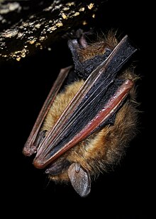


Species of bat Tricolored bat A tricolored bat captured near Arnold Air Force Base in 2022 Conservation status Vulnerable  (IUCN 3.1)[1] Scientific classification Domain: Eukaryota Kingdom: Animalia Phylum: Chordata Class: Mammalia Order: Chiroptera Family: Vespertilionidae Tribe: Perimyotini Genus: PerimyotisMenu, 1984 Species: P. subflavus Binomial name Perimyotis subflavus(F. Cuvier, 1832) Range of tricolored bat (in yellow) Synonyms Vespertilio subflavus F. Cuvier, 1832 ...


Dominican baseball player (born 1998) Baseball player Joan AdonAdon with the Nationals in 2022Washington Nationals – No. 60PitcherBorn: (1998-08-12) August 12, 1998 (age 25)Santo Domingo, Dominican RepublicBats: RightThrows: RightMLB debutOctober 3, 2021, for the Washington NationalsMLB statistics (through April 9, 2024)Win–loss record3–16Earned run average6.52Strikeouts114 Teams Washington Nationals (2021–present) Joan Manuel Adon (born August 12, 1998) is a Domin...

Artikel utama: Piala Dunia FIFA 2010 dan Babak penyisihan grup Piala Dunia FIFA 2010 Pertandingan di Grup A Piala Dunia FIFA 2010 akan diadakan dari tanggal 11 Juni sampai dengan 22 Juni 2010.[1] Grup ini terdiri dari tuan rumah Afrika Selatan, Meksiko, Uruguay, dan Prancis. Prancis dan Uruguay pernah bertemu pada Piala Dunia FIFA 2002 dimana pertandingan berakhir sengan skor 0-0. Pemenang grup ini yaitu, Uruguay, melaju melawan runner-up Grup B yaitu Korea Selatan di babak gugur. Run...


English footballer Paul Tomlinson Personal informationFull name Paul Tomlinson[1]Date of birth (1965-02-04) 4 February 1965 (age 59)[2]Place of birth Rotherham,[2] EnglandHeight 6 ft 2 in (1.88 m)[3]Position(s) GoalkeeperSenior career*Years Team Apps (Gls)1983–1987 Sheffield United 37 (0)1986–1987 → Birmingham City (loan) 11 (0)1987–1995 Bradford City 293 (0)Total 341 (0) *Club domestic league appearances and goals Paul Tomlinson (bo...


American football player and coach (1920–1983) American football player Bob WaterfieldWaterfield c. 1947No. 7Position:Quarterback Safety Kicker PunterPersonal informationBorn:(1920-07-26)July 26, 1920Elmira, New York, U.S.Died:March 25, 1983(1983-03-25) (aged 62)Burbank, California, U.S.[1]Height:6 ft 1 in (1.85 m)Weight:200 lb (91 kg)Career informationHigh school:Van Nuys (Los Angeles, California)College:UCLA (1941–1942, 1944)NFL draft:1944 / Ro...

Type of girder The old Britannia Bridge with train track inside the box-girder tunnel. Section of the original tubular Britannia Bridge The patent curved and tapered box girder jib of a Fairbairn steam crane A box girder or tubular girder (or box beam) is a girder that forms an enclosed tube with multiple walls, as opposed to an I- or H-beam. Originally constructed of wrought iron joined by riveting, they are now made of rolled or welded steel, aluminium extrusions or prestressed concrete. Co...


Untuk gubernur pra-Mongol Hulagu di Persia pada pertengahan abad ke-13, lihat Arghun Aqa. Untuk desa di Iran, lihat Arghun, Iran. Untuk kota di Afghanistan, lihat Urgun. Arghun ᠠᠷᠭᠤᠨKhanSebuah lukisan yang menggambarkan Arghun (berdiri, menggendong putranya Ghazan) di bawah payung kerajaan. Di sebelahnya adalah ayahnya Abaqa duduk di atas kuda.Berkuasa1284–7 Maret 1291PendahuluTekuderPenerusGaykhatuAyahAbaqaPasanganQuthluq KhatunUruk KhatunTodai KhatunSaljuk KhatunBulughan KhatunQ...


Biografi tokoh yang masih hidup ini tidak memiliki referensi atau sumber sehingga isinya tidak dapat dipastikan. Bantu memperbaiki artikel ini dengan menambahkan sumber tepercaya. Materi kontroversial atau trivial yang sumbernya tidak memadai atau tidak bisa dipercaya harus segera dihapus.Cari sumber: Meivin Malelak – berita · surat kabar · buku · cendekiawan · JSTOR (Pelajari cara dan kapan saatnya untuk menghapus pesan templat ini) Meivin MalelakLahi...

Medical school in St. Louis, Missouri, US Washington University School of MedicineTypePrivateEstablished1891Parent institutionWashington University in St. LouisDeanDavid PerlmutterAcademic staff1874Students1349 (including 605 MD [183 MD/PhD] and 267 OT, 278 PT)LocationSt. Louis, Missouri, United StatesCampusUrbanWebsitemedicine.wustl.edu BJC Institute of Health on the Washington University School of Medicine campus Washington University School of Medicine (WUSM) is the medical school of Washi...


Periodic comet with 12 year orbit 92P/SanguinDiscoveryDiscovered byJuan G. SanguinDiscovery dateOctober 15, 1977Orbital characteristicsEpochNovember 17, 2014Observation arc36 yearsNumber ofobservations896Aphelion8.89 AUPerihelion1.82 AUSemi-major axis5.36 AUEccentricity.659521857266038Orbital period12 yearsInclination19.44339335368171Last perihelion2015-Mar-012002-Sep-23Next perihelion2027-July-15[1]TJupiter2.410 Physical characteristicsDimensions2.38 kmComet totalmagnitude ...


Questa voce sull'argomento ginnasti statunitensi è solo un abbozzo. Contribuisci a migliorarla secondo le convenzioni di Wikipedia. Theodore StudierNazionalità Stati Uniti Ginnastica SocietàTurnverein Vorwärts   Modifica dati su Wikidata · Manuale Theodore Frederick Studier (Ohio, 11 maggio 1882 – Cleveland, 14 marzo 1959) è stato un ginnasta statunitense. Carriera Studier partecipò ai Giochi olimpici di Saint Louis 1904 nelle gare di ginnastica. Giunse tredicesi...

Bagian dari seriGenetika   Komponen penting Kromosom DNA RNA Genom Pewarisan Mutasi Nukleotida Variasi Garis besar Indeks Sejarah dan topik Pengantar Sejarah Evolusi (molekuler) Genetika populasi Hukum Pewarisan Mendel Genetika kuantitatif Genetika molekuler Penelitan Pengurutan DNA Rekayasa genetika Genomika ( templat) Genetika medis Cabang-cabang genetika Pengobatan personal Pengobatan personal lbs Evolusi molekuler merupakan proses evolusi yang terjadi pada skala DNA, RNA, dan protein...


Der Titel dieses Artikels ist mehrdeutig. Weitere Bedeutungen sind unter Literarischer Salon (Begriffsklärung) aufgeführt. Der literarische Salon von Madame Geoffrin (1755) Ein literarischer Salon war ein zumeist privater gesellschaftlicher Treffpunkt für Diskussionen, Lesungen oder musikalische Veranstaltungen vom 17. bis zum 20. Jahrhundert. Neben literarischen und sonstigen künstlerischen Salons gab es auch politische (Spitzemberg, Treuberg) und wissenschaftliche (Helmholtz) Salo...


American philosopher This biography of a living person needs additional citations for verification. Please help by adding reliable sources. Contentious material about living persons that is unsourced or poorly sourced must be removed immediately from the article and its talk page, especially if potentially libelous.Find sources: William Hirstein – news · newspapers · books · scholar · JSTOR (March 2024) (Learn how and when to remove this message) Willi...

يفتقر محتوى هذه المقالة إلى الاستشهاد بمصادر. فضلاً، ساهم في تطوير هذه المقالة من خلال إضافة مصادر موثوق بها. أي معلومات غير موثقة يمكن التشكيك بها وإزالتها. (سبتمبر 2023) NGC 715   الكوكبة قيطس  رمز الفهرس 2MASX J01531252-1252221 (Two Micron All-Sky Survey, Extended source catalogue)6dFGS gJ015312.5-125222 (6dF Galaxy Survey)GSC 0...


TG LA7PaeseItalia Anno2001 – in produzione Generetelegiornale Durata10 minuti (edizione delle 7:40)45 minuti (edizione delle 13:30)5 minuti (edizione delle 18:15)40 minuti (edizione delle 20:00)10 minuti (edizione della notte) Lingua originaleitaliano RealizzazioneConduttorevari Regiavari MusicheSilvio Amato Casa di produzioneLA7 S.r.l. Rete televisivaLA7LA7d Modifica dati su Wikidata · Manuale Il TG LA7 è il telegiornale di LA7. L'attuale direttore è Enrico Mentana. Indice 1 S...