Eye development

Transverse section of head of chick embryo of forty-eight hours’ incubation
Transverse section of head of chick embryo of fifty-two hours’ incubation, showing the lens and the optic cup

Eye formation in the human embryo begins at approximately three weeks into embryonic development and continues through the tenth week.[1] Cells from both the mesodermal and the ectodermal tissues contribute to the formation of the eye. Specifically, the eye is derived from the neuroepithelium, surface ectoderm, and the extracellular mesenchyme which consists of both the neural crest and mesoderm.[2][3][4]

Neuroepithelium forms the retina, ciliary body, iris, and optic nerves. Surface ectoderm forms the lens, corneal epithelium and eyelid. The extracellular mesenchyme forms the sclera, the corneal endothelium and stroma, blood vessels, muscles, and vitreous.

The eye begins to develop as a pair of optic vesicles on each side of the forebrain at the end of the fourth week of pregnancy. Optic vesicles are outgrowings of the brain which make contact with the surface ectoderm and this contact induces changes necessary for further development of the eye. Through a groove at the bottom of the optic vesicle known as choroid fissure the blood vessels enter the eye. Several layers such as the neural tube, neural crest, surface ectoderm, and mesoderm contribute to the development of the eye.[2][3][4]

Eye development is initiated by the master control gene PAX6, a homeobox gene with known homologues in humans (aniridia), mice (small eye), and Drosophila (eyeless). The PAX6 gene locus is a transcription factor for the various genes and growth factors involved in eye formation.[1][5] Eye morphogenesis begins with the evagination, or outgrowth, of the optic grooves or sulci. These two grooves in the neural folds transform into optic vesicles with the closure of the neural tube.[6] The optic vesicles then develop into the optic cup with the inner layer forming the retina and the outer portion forming the retinal pigment epithelium. The middle portion of the optic cup develops into the ciliary body and iris.[7] During the invagination of the optic cup, the ectoderm begins to thicken and form the lens placode, which eventually separates from the ectoderm to form the lens vesicle at the open end of the optic cup.[1][3][4]

Further differentiation and mechanical rearrangement of cells in and around the optic cup gives rise to the fully developed eye.

Sequential inductions

This development is an example of sequential inductions where the organ is formed from three different tissues:

Neural tube ectoderm (neuroectoderm)

First, there is an outpocketing of the neural tube called optic vesicles. Development of the optic vesicles starts in the 3-week embryo, from a progressively deepening groove in the neural plate called the optic sulcus. Some studies suggest this mechanism is regulated by RX/RAX transcription factor.[8] The proteins Wnt and FGF (fibroblast growth factor) play a part in this early stage and are regulated by another protein called Shisa.[6] As this expands, the rostral neuropore (the exit of the brain cavity out of the embryo) closes and the optic sulcus and the neural plate becomes the optic vesicle.[9] Optic nerves arise from connections of the vesicles to the forebrain.[1]

Neuroectoderm gives rise to the following compartments of the eye:

Surface ectoderm

Lens development is closely related to optic vesicle development. The interaction between the growing vesicle and the ectoderm causes the ectoderm to thicken at that point. This thickened portion of the ectoderm is called the lens placode. Next, the placode invaginates and forms a pouch referred to as the lens pit.[1][3][4] Scientists are studying the tension forces necessary for invagination of the lens placode and current research suggests that microfilaments might be present in early retinal cells to allow for invagination behavior. Research has also shown that Rho GTPase dependent filopodia from the precursor lens ectoderm play an important role in the formation of the lens pit.[10][3][4] Eventually, the pit becomes completely enclosed. This enclosed structure is the lens vesicle.[1] Studies have shown that lens development requires the presence of the Pax6 gene, which is the master regulatory gene for eye morphogenesis.[11] This master regulatory gene is not necessary for the closely associated optic vesicle development.[12] Additionally, Ras activation has been shown to be sufficient for starting lens differentiation, but not enough for its completion.[11]

The optic vesicles then begin to form the optic cup.[3][4] Optic cup morphogenesis is the invagination process occurring after neuroectoderm movement forms the spherical optic vesicle (Phase 1). Invagination is when a tissue folds back on itself. Over the course of approximately 12 hours, the distal end of the optic vesicle inner layer begins to flatten (Phase 2). Over the following 18 hours, both the inner and outer layers begin to flex inward at sharp angles, beginning the formation of a C-shaped edge (Phase 3). The final 18 hours involve continuing this apically convex invagination to form the optic cup [3] [4] . At this point, morphologies such as columnar epithelial cells, pseudo-stratified cells, and apically narrow wedge-shaped cells can be observed.[13]

The inner layer of the optic cup is made of neuroepithelium (neural retina), while the outer layer is composed of retinal pigment epithelium (RPE). Experiments have determined that RPE cell differentiation and maintenance requires interaction with neighboring tissues, most likely canonical Wnt signaling, while neural retina differentiation is driven by tissue-autonomous factors.[13]

Bone morphogenic proteins (BMPs) are important regulators of optic cup development. In fact, research studies have shown that BMP agonists and antagonists are necessary for precision of optic cup development.[12] Interactions between tissues and signaling pathways also play a major role in morphogenesis of the optic cup.[8]

It is of interest to note that research has shown isolating the optic cup from neighboring tissue after completed invagination in tissue culture medium can lead to the development of most major parts of the eye, including photoreceptors, ganglion cells, bipolar cells, horizontal cells, amacrine cells and Muller glia. This indicates that morphogenesis of the optic cup occurs independently of external cues from its environment, including presence of lens.[13] However, the lens is necessary to act as an inducer for the ectoderm to transform it into the cornea.

Surface ectoderm produces the following parts:

Neural crest

Neural crest cells are themselves derived from the ectoderm and lie close to the neural tube:

Mesoderm

Mesoderm contributes to the following structures:

Developmental cascade

According to Liem et al., the organogenesis of the eye is pointed out as an example of a developmental cascade of inductions. The eye is essentially a derivative of the ectoderm from the somatic ectoderm and neural tube, with a succession of inductions by the chordamesoderm.

Chordamesoderm induces the anterior portion of the neural tube to form the precursors of the synapomorphic tripartite brain of vertebrates, and it will form a bulge called the diencephalon. Further induction by the chordamesoderm will form a protrusion: the optic vesicle. This vesicle will be subsequently invaginated by means of further inductions from the chordamesoderm. The optic vesicle will then induce the ectoderm that thickens (lens placode) and further invaginates to a point that detaches from the ectoderm and forms a neurogenic placode by itself. The lens placode is affected by the chordamesoderm making it invaginate and forms the optic cup composed by an inner layer of the neural retina and outer layer of the pigmented retina that will unite and form the optic stalk. The pigmented retina is formed by rods and cones and composed of small cilia typical of the ependymal epithelium of the neural tube. Some cells in the lens vesicle will be fated to form the cornea and the lens vesicle will develop completely to form the definitive lens. Iris is formed from the optic cup cells.

Responsivity of head epidermis

Only the epidermis in the head is competent to respond to the signal from the optic vesicles. Both the optic vesicle and the head epidermis are required for eye development. The competence of the head epidermis to respond to the optic vesicle signals comes from the expression of Pax6 in the epidermis. Pax6 is necessary and sufficient for eye induction. This competence is acquired gradually during gastrulation and neurulation from interactions with the endoderm, mesoderm, and neural plate.

Regulation and inhibition

Sonic hedgehog reduces the expression of Pax6. When Shh is inhibited during development, the domain of expression for Pax6 is expanded and the eyes fail to separate causing cyclopia.[14] Overexpression of Shh causes a loss of eye structures.

Retinoic acid generated from vitamin A in the retina plays an essential role in eye development as a secreted paracrine signal which restricts invasion of perioptic mesenchyme around the optic cup.[15] Vitamin A deficiency during embryogenesis results in anterior segment defects (particularly cornea and eyelids) that lead to vision loss or blindness.

There is some evidence that LMX1B plays a role in periocular mesenchymal survival.[16]

Additional images

References

  1. ^ a b c d e f Ort, D., David, H., "Development of the Eye". Retrieved 22 April 2015.
  2. ^ a b Sadler, T.W (1990). Langman's medical embryology (6th ed.). Williams and Wilkins. ISBN 978-0683074932.
  3. ^ a b c d e f g Hosseini, Hadi S.; Beebe, David C.; Taber, Larry A. (2014). "Mechanical effects of the surface ectoderm on optic vesicle morphogenesis in the chick embryo". Journal of Biomechanics. 47 (16): 3837–3846. doi:10.1016/j.jbiomech.2014.10.018. PMC 4261019. PMID 25458577.
  4. ^ a b c d e f g Hosseini, Hadi S.; Taber, Larry A. (2018). "How mechanical forces shape the developing eye". Progress in Biophysics and Molecular Biology. 137 (16): 25–36. doi:10.1016/j.pbiomolbio.2018.01.004. PMC 6085168. PMID 29432780.
  5. ^ Keller, A. M. V., "Embryonic Development of the Eye" Archived 2015-04-08 at the Wayback Machine. Retrieved 22 April 2015.
  6. ^ a b Fuhrmann, S., Levine, E. M. and Reh, T. A. (2000). "Extraocular mesenchyme patterns the optic vesicle during early eye development in the embryonic chick". Development 127, 4599–4609.
  7. ^ LifeMap Science, Inc. "Embryonic and Postnatal Development of the Eye". Retrieved 22 April 2015.
  8. ^ a b Fuhrmann, S., “Eye Morphogenesis and Patterning of the Optic Vesicle” Current Topics in Developmental Biology 93, 61-84 (07 October 2010)
  9. ^ "eye(vertebrate) McGraw-Hil; Encyclopedia of Science and Technology (2007), vol.6, pp.801-802
  10. ^ Fuhrmann, Sabine, “Eye Morphogenesis and Patterning of the Optic Vesicle” Current Topics in Developmental Biology 93, 61-84 (07 October 2010)
  11. ^ a b Hill, M.A. (2015) Embryology Vision - Lens Development. Retrieved April 22, 2015.
  12. ^ a b Adler, R., Canto-Soler, M.V., “Molecular mechanisms of optic vesicle development: Complexities, ambiguities and controversies”, Developmental Biology 305,1,1-13 (1 May 2007)
  13. ^ a b c Eiraku et al., "Self-organizing optic-cup morphogenesis in three-dimensional cell culture", Nature 472, 51–56 (07 April 2011)
  14. ^ "Development of the Vertebrate Eye -- Developmental Biology -- NCBI Bookshelf". Retrieved 2010-04-10.
  15. ^ Duester, G (September 2008). "Retinoic acid synthesis and signaling during early organogenesis". Cell. 134 (6): 921–31. doi:10.1016/j.cell.2008.09.002. PMC 2632951. PMID 18805086.
  16. ^ McMahon C, Gestri G, Wilson SW, Link BA (August 2009). "Lmx1b is essential for survival of periocular mesenchymal cells and influences Fgf-mediated retinal patterning in zebrafish". Dev. Biol. 332 (2): 287–98. doi:10.1016/j.ydbio.2009.05.577. PMC 2716413. PMID 19500562.

Further reading

  • Keith L. Moore and T.V.N. Persaud (2008). The developing human—clinical oriented embryology. 8th edition. USA: Saunders, an imprint of Elsevier Inc. p. 429

Read other articles:

Artikel ini sebatang kara, artinya tidak ada artikel lain yang memiliki pranala balik ke halaman ini.Bantulah menambah pranala ke artikel ini dari artikel yang berhubungan atau coba peralatan pencari pranala.Tag ini diberikan pada Desember 2023. A.M. Tambunan dapat mengacu pada beberapa hal berikut: Albert Mangaratua Tambunan (1912 – 1970) politikus Parkindo, mantan Menteri Sosial Indonesia Albertus Maruli Tambunan (1925 – 2019) perwira tinggi angkatan darat Indo...


Kotak rangkap generik Kotak rangkap adalah gawit (atau kontrol) antarmuka pengguna grafis yang umum digunakan. Secara tradisional, ini adalah kombinasi daftar naik-turun atau kotak daftar dan kotak teks satu baris yang dapat disuntin, memungkinkan pengguna mengetikkan nilai secara langsung atau memilih nilai dari daftar. Istilah kotak rangkap kadang-kadang digunakan untuk mengartikan daftar naik-turun. [1] Baik di Java maupun . NET, kotak rangkap bukan sinonim untuk daftar naik-turun....
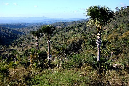
 

9°41′41″S 124°42′11″E / 9.69472°S 124.70306°E / -9.69472; 124.70306 Amanatun UtaraKecamatanNegara IndonesiaProvinsiNusa Tenggara TimurKabupatenTimor Tengah SelatanPemerintahan • Camat-Populasi • Total- jiwaKode Kemendagri53.02.09 Kode BPS5304110 Luas- km²Desa/kelurahan- Savana berbukit dekat Ayotupas Amanatun Utara adalah sebuah kecamatan di Kabupaten Timor Tengah Selatan, Nusa Tenggara Timur, Indonesia. Kecamatan ini beribu ko...

Strada statale 513di Val d'EnzaLocalizzazioneStato Italia Regioni Emilia-Romagna DatiClassificazioneStrada statale InizioParma FineCastelnovo ne' Monti Lunghezza56,270[1] km Provvedimento di istituzioneD.M. 27/12/1966 - G.U. 34 dell'8/02/1967[2] GestoreTratte ANAS: nessuna (dal 2001 la gestione è passata alla Provincia di Parma e alla Provincia di Reggio Emilia) Manuale La ex strada statale 513 di Val d'Enza (SS 513), ora strada provinciale 513 R di Val d'Enza (SP 5...


Universitas Prasetiya MulyaMotoA COLLABORATIVE LEARNING BY ENTERPRISINGJenisUniversitas swastaDidirikan1982RektorProf. Dr. Djisman S. Simandjuntak DekanDr. Fathony Rahman Dr. N. Hassan Wirajuda Stevanus Wisnu Wijaya, Ph.D.AlamatKavling Edutown I.1 Jl. BSD Raya Utama, BSD City, Tangerang, IndonesiaWarnaBiru TuaNama julukanPrasmulyanSitus webwww.prasetiyamulya.ac.id Universitas Prasetiya Mulya adalah institusi pendidikan di Indonesia yang berbasis di BSD City, Banten. Universitas ini memiliki k...
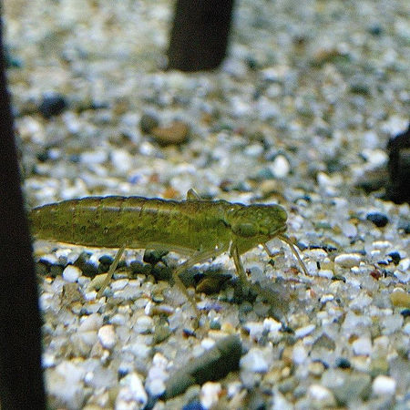
 

Capung Anisoptera Yellow-winged darter (en) TaksonomiKerajaanAnimaliaFilumArthropodaKelasInsectaOrdoOdonataUpaordoAnisoptera Selys, 1854 lbs Capung atau sibar-sibar adalah kelompok serangga yang tergolong ke dalam bangsa Odonata. Kedua macam serangga ini jarang berada jauh-jauh dari air, tempat mereka bertelur dan menghabiskan masa pra-dewasa anak-anaknya. Namanya dalam bahasa daerah adalah papatong (Sd.), kinjeng (Jw.), coblang (Jw.), kasasiur (bjn), tjapung, Sansibur (DykNgj) Bungkoloko (Si...

Langeland ialah pulau di Denmark yang terletak antara Sabuk Besar dan Teluk Kiel. Memiliki luas 285 km² (skt. 110 mil persegi), dan berpenduduk sekitar 15.000. Pulau ini memproduksi padi dan dikenal sebagai objek wisata. Sebuah jembatan menghubungkannya ke Taasinge dan Fyn. Secara administratif, pulau ini dibagi atas 3 kotamadya: Rudkøbing, Sydlangeland, dan Tranekær. Semuanya akan bergabung pada 1 Januari 2007 sebagai bagian dari reformasi skala nasional dan membentuk kotamadya Lange...


Административное деление Италии Топонимия Италии — совокупность географических названий, включающая наименования природных и культурных объектов на территории Италии. Структура и состав топонимии страны обусловлены её географическим положением, богатой историей...


ColumbiformesRentang fosil: Miosen Awal - Sekarang Tekukur Biasa Klasifikasi ilmiah Kerajaan: Animalia Filum: Chordata Kelas: Aves Infrakelas: Neognathae Ordo: ColumbiformesLatham, 1790 Famili Columbidae Raphidae Ordo burung Columbiformes mencakup dara dan merpati yang tersebar luas di seluruh penjuru dunia dan diklasifikasikan ke dalam famili Columbidae, dan Dodo & Solitaire Rodrigues yang telah punah dan telah lama diklasifikasikan ke dalam famili kedua, Raphidae.[1] 313 spesie...

2006 protest This article needs additional citations for verification. Please help improve this article by adding citations to reliable sources. Unsourced material may be challenged and removed.Find sources: Great American Boycott – news · newspapers · books · scholar · JSTOR (May 2009) (Learn how and when to remove this template message) The Great American BoycottDateMay 1, 2006 (2006-05-01)LocationUnited States, nationwideTypeDemonstra...
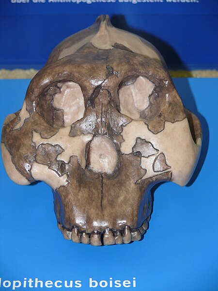
 

Robust australopithecinesRentang fosil: Pleistosen Skull of Paranthropus boisei Klasifikasi ilmiah Kerajaan: Animalia Filum: Chordata Kelas: Mammalia Ordo: Primates Famili: Hominidae Subfamili: Homininae Tribus: Hominini Subtribus: Hominina Genus: ParanthropusBroom, 1938 Species †Paranthropus aethiopicus †Paranthropus boisei †Paranthropus robustus Robust australopithecines, anggota genus hominin Paranthropus yang telah punah, adalah hominin bipedal yang merupakan keturunan dari hominin...


Muhammad Taufiq Arasj Waasops Panglima TNIPetahanaMulai menjabat 1 April 2024PendahuluFebriel Buyung SikumbangPenggantiPetahanaKomandan Lanud Atang SendjajaMasa jabatan29 Maret 2023 – 1 April 2024PendahuluSulionoPenggantiJuli Heryanto Ginting Informasi pribadiLahir6 Agustus 1973 (umur 50)Ujung Pandang, Sulawesi SelatanKebangsaanIndonesiaAlma materAkademi Angkatan Udara (1995)Karier militerPihak IndonesiaDinas/cabang TNI Angkatan UdaraMasa dinas1995—sekarangPang...

Galaxy containing well-studied supermassive black hole IRAS 13224-3809Observation data (J2000[1] epoch)ConstellationCentaurus[2]Right ascension13h 25m 19.38s[1]Declination−38° 24′ 52.61″[1]Redshift0.06580 ± 0.00018Distance1 billion light-years[3]Apparent magnitude (V)13.80[4]Other designations2MASX J13251937-3824524; 2MASS J13251937-3824526; GSC 07787-00931; IRAS F13224-3809; 1RXS J132519.4-382445; WISE J132519...


Atlanta BravesBaseball Campione della NL East division in carica Segni distintivi Uniformi di gara Casa Trasferta Terza Colori sociali               Blu marino, rosso scarlatto, bianco Dati societari Città Atlanta Nazione  Stati Uniti Lega National League Division East Fondazione 1871 Denominazione Boston Red Stockings(1871-1876)Boston Red Caps(1876-1882)Boston Beaneaters(1883-1906)Boston Doves(1907-1910)Boston Rustlers(1911)B...


Halaman ini berisi artikel tentang kabupaten di Indonesia. Untuk kota bernama sama, lihat Kota Serang. Untuk kegunaan lain, lihat Serang (disambiguasi). Kabupaten SerangKabupatenTranskripsi bahasa daerah • Aksara Sundaᮞᮦᮛᮀ • Cacarakanꦱꦺꦫꦁ • PegonسيراڠPemandangan di Pantai Anyer. LambangMotto: Sepi ing pamrih, rame ing gawe(Jawa) Bekerja tanpa mengharap imbalan apa punPetaKabupaten SerangPetaTampilkan peta JawaKabupaten SerangK...

Disused railway station in North Yorkshire, England BeckholeBeckhole station site 1988General informationLocationBeck Hole, ScarboroughEnglandCoordinates54°24′28″N 0°44′11″W / 54.407889°N 0.736475°W / 54.407889; -0.736475Grid referenceNZ821020Other informationStatusDisusedHistoryOriginal companyWhitby and Pickering RailwayPre-groupingNorth Eastern RailwayKey dates1836OpenedClosed to passengers1 July 1865Reopened for passengers (summers only)19081914Closed f...


This article is about the administrative county. For the Riksdag constituency, see Uppsala County (Riksdag constituency). County (län) of Sweden County of SwedenUppsala County Uppsala län (Swedish)County of Sweden FlagCoat of armsUppsala County in SwedenLocation map of Uppsala County in SwedenCoordinates: 59°51′30″N 17°39′00″E / 59.85833°N 17.65000°E / 59.85833; 17.65000CountrySwedenFormed1634CapitalUppsalaMunicipalities 8 ÄlvkarlebyEnköpingHåboHe...


Study and practice to minimize the occurrence and consequences of motor vehicle accidents Passive safety and ETSC redirect here. For nuclear safety, see Passive nuclear safety. For the former East Texas State College, see Texas A&M University–Commerce. Passive restraint redirects here. For the Clutch EP, see Passive Restraints. Crash testing is one of the components of automotive safety. Automotive safety is the study and practice of automotive design, construction, equipment and regula...

لويس الحادي عشر ملك فرنسا (بالفرنسية: Louis XI)‏  معلومات شخصية الميلاد 3 يوليو 1423(1423-07-03)بورجيز الوفاة 30 أغسطس 1483 (60 سنة) سبب الوفاة سكتة دماغية  مواطنة فرنسا  الديانة الكنيسة الرومانية الكاثوليكية الزوجة مارغريت ستيوارت (24 يونيو 1436–16 أغسطس 1445)شارلوت من سافوي (9 مارس 1451–...


Filmfare Award for Best Actor2022 winner: Biju Menon for Ayyappanum KoshiyumAwarded forBest Performance by an Actor in a Leading Role in Malayalam FilmsCountryIndiaPresented byFilmfareFirst awardedMadhu,Swayamvaram (1972)Currently held byBiju Menon, Ayyappanum Koshiyum (2022)HighlightsMost awardsMammootty (12)Total awarded48First winnerMadhuWebsitefilmfare.com The Filmfare Award for Best Actor – Malayalam is an award instituted in 1972, presently annually at the Filmfare Awards South to an...