Seminal vesicles

Seminal vesicle
Cross-section of the lower abdomen in a male, showing parts of the urinary tract and male reproductive system, with the seminal vesicles seen top right
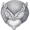
The seminal vesicles seen near the prostate, viewed from in front and above.
Details
PrecursorMesonephric ducts
SystemMale reproductive system
ArteryInferior vesical artery, middle rectal artery
LymphExternal iliac lymph nodes, internal iliac lymph nodes
Identifiers
Latinvesiculae seminales, glandulae vesiculosae
MeSHD012669
TA98A09.3.06.001
TA23631
FMA19386
Anatomical terminology

The seminal vesicles (also called vesicular glands[1] or seminal glands) are a pair of convoluted tubular accessory glands that lie behind the urinary bladder of male mammals. They secrete fluid that largely composes the semen.

The vesicles are 5–10 cm in size, 3–5 cm in diameter, and are located between the bladder and the rectum. They have multiple outpouchings, which contain secretory glands, which join together with the vasa deferentia at the ejaculatory ducts. They receive blood from the vesiculodeferential artery, and drain into the vesiculodeferential veins. The glands are lined with column-shaped and cuboidal cells. The vesicles are present in many groups of mammals, but not marsupials, monotremes or carnivores.

Inflammation of the seminal vesicles is called seminal vesiculitis and most often is due to bacterial infection as a result of a sexually transmitted infection or following a surgical procedure. Seminal vesiculitis can cause pain in the lower abdomen, scrotum, penis or peritoneum, painful ejaculation, and blood in the semen. It is usually treated with antibiotics, although may require surgical drainage in complicated cases. Other conditions may affect the vesicles, including congenital abnormalities such as failure or incomplete formation, and, uncommonly, tumours.

The seminal vesicles have been described as early as the second century AD by Galen, although the vesicles only received their name much later, as they were initially described using the term from which the word prostate is derived.

Structure

The human seminal vesicles are a pair of glands in males that are positioned below the urinary bladder and at the end of the vasa deferentia, where they enter the prostate. Each vesicle is a coiled and folded tube, with occasional outpouchings termed diverticula in its wall.[2] The lower part of the tube ends as a straight tube called the excretory duct, which joins with the vas deferens of that side of the body to form an ejaculatory duct. The ejaculatory ducts pass through the prostate gland before opening separately into the verumontanum of the prostatic urethra.[2] The vesicles are between 5–10 cm in size, 3–5 cm in diameter, and have a volume of around 13 mL.[3]

The vesicles receive blood supply from the vesiculodeferential artery, and also from the inferior vesical artery. The vesiculodeferential artery arises from the umbilical arteries, which branch directly from the internal iliac arteries.[3] Blood is drained into the vesiculodeferential veins and the inferior vesical plexus, which drain into the internal iliac veins.[3] Lymphatic drainage occurs along the venous routes, draining into the internal iliac nodes.[3]

The vesicles lie behind the bladder at the end of the vasa deferentia. They lie in the space between the bladder and the rectum; the bladder and prostate lie in front, the tip of the ureter as it enters the bladder above, and Denonvilliers' fascia and the rectum behind.[3]

Development

In the developing embryo, at the hind end lies a cloaca. This, over the fourth to the seventh week, divides into a urogenital sinus and the beginnings of the anal canal, with a wall forming between these two inpouchings called the urorectal septum.[4] Two ducts form next to each other that connect to the urogenital sinus; the mesonephric duct and the paramesonephric duct, which go on to form the reproductive tracts of the male and female respectively.[4]

In the male, under the influence of testosterone, the mesonephric ducts proliferate, forming the epididymis, ductus deferens and, via a small outpouching near the developing prostate, the seminal vesicles.[4] Sertoli cells secrete anti-Müllerian hormone, which causes the paramesonephric ducts to regress.[4]

The development and maintenance of the seminal vesicles, as well as their secretion and size/weight, are highly dependent on androgens.[5][6] The seminal vesicles contain 5α-reductase, which metabolizes testosterone into its much more potent metabolite, dihydrotestosterone (DHT).[6] The seminal vesicles have also been found to contain luteinizing hormone receptors, and hence may also be regulated by the ligand of this receptor, luteinizing hormone.[6]

Microanatomy

Histology of seminal vesicle glands on H&E stain. They may mimic prostatic adenocarcinoma by crowded glands with enlarged hyperchromatic and irregular nuclei, but will have inconspicuous nucleoli and coarse refractile golden brown lipofuscin granules.[7]

The inner lining of the seminal vesicles (the epithelium) is made of a lining of interspersed column-shaped and cube-shaped cells.[8] There are varying descriptions of the lining as being pseudostratified and consisting of column-shaped cells only.[9] When viewed under a microscope, the cells are seen to have large bubbles in their interior. This is because their interior, called cytoplasm, contains lipid droplets involved in secretion during ejaculation.[8] The tissue of the seminal vesicles is full of glands, spaced irregularly.[8] As well as glands, the seminal vesicles contain smooth muscle and connective tissue.[8] This fibrous and muscular tissue surrounds the glands, helping to expel their contents.[3] The outer surface of the glands is covered in peritoneum.[3]

Function

The seminal vesicles secrete a significant proportion of the fluid that ultimately becomes semen.[10] Fluid is secreted from the ejaculatory ducts of the vesicles into the vas deferens, where it becomes part of semen. This then passes through the urethra, where it is ejaculated during a male sexual response.[9]

About 70-85% of the seminal fluid in humans originates from the seminal vesicles.[11] The fluid consists of nutrients including fructose and citric acid, prostaglandins, and fibrinogen.[10] Fructose is not produced anywhere else in the body except in the seminal vesicles. It provides a forensic test in rape cases.

Nutrients help support sperm until fertilisation occurs; prostaglandins may also assist by softening mucus of the cervix, and by causing reverse contractions of parts of the female reproductive tract such as the fallopian tubes, to ensure that sperm are less likely to be expelled.[10]

Clinical significance

Disease

Diseases of the seminal vesicles as opposed to that of prostate gland are extremely rare and are infrequently reported in the medical literature.[12]

Congenital anomalies associated with the seminal vesicles include failure to develop, either completely (agenesis) or partially (hypoplasia), and cysts.[13][14] Failure of the vesicles to form is often associated with absent vas deferens, or an abnormal connection between the vas deferens and the ureter.[3] The seminal vesicles may also be affected by cysts, amyloidosis, and stones.[13][14] Stones or cysts that become infected, or obstruct the vas deferens or seminal vesicles, may require surgical intervention.[9]

Seminal vesiculitis (also known as spermatocystitis) is an inflammation of the seminal vesicles, most often caused by bacterial infection.[15] Symptoms can include vague back or lower abdominal pain; pain of the penis, scrotum or peritoneum; painful ejaculation; blood in the semen on ejaculation; irritative and obstructive voiding symptoms; and impotence.[16] Infection may be due to sexually transmitted infections, as a complication of a procedure such as prostate biopsy.[9] It is usually treated with antibiotics. If a person experiences ongoing discomfort, transurethral seminal vesiculoscopy may be considered.[17][18] Intervention in the form of drainage through the skin or surgery may also be required if the infection becomes an abscess.[9] The seminal vesicles may also be affected by tuberculosis, schistosomiasis and hydatid disease.[13][14] These diseases are investigated, diagnosed and treated according to the underlying disease.[9]

Benign tumours of the seminal vesicles are rare.[9] When they do occur, they are usually papillary adenomata and cystadenomata. They do not cause elevation of tumour markers and are usually diagnosed based on examination of tissue that has been removed after surgery.[9] Primary adenocarcinoma, although rare, constitutes the most common malignant tumour of the seminal vesicles;[19] that said, malignant involvement of the vesicles is typically the result of local invasion from an extra-vesicular lesion.[9] When adenocarcinoma occurs, it can cause blood in the urine, blood in the semen, painful urination, urinary retention, or even urinary obstruction.[9] Adenocarcinomata are usually diagnosed after they are excised, based on tissue diagnosis.[9] Some produce the tumour marker Ca-125, which can be used to monitor for reoccurence afterwards.[9] Even rarer neoplasms include sarcoma, squamous cell carcinoma, yolk sac tumour, neuroendocrine carcinoma, paraganglioma, epithelial stromal tumours and lymphoma.[19]

Investigations

Symptoms due to diseases of the seminal vesicles may be vague and not able to be specifically attributable to the vesicles themselves; additionally, some conditions such as tumours or cysts may not cause any symptoms at all.[9] When diseases is suspected, such as due to pain on ejaculation, blood in the urine, infertility, due to urinary tract obstruction, further investigations may be conducted.[9]

A digital rectal examination, which involves a finger inserted by a medical practitioner through the anus, may cause greater than usual tenderness of the prostate gland, or may reveal a large seminal vesicle.[9] Palpation is dependent on the length of index finger as seminal vesicles are located above the prostate gland and retrovesical (behind the bladder).

A urine specimen may be collected, and is likely to demonstrate blood within the urine.[9] Laboratory examination of seminal vesicle fluid requires a semen sample, e.g. for semen culture or semen analysis. Fructose levels provide a measure of seminal vesicle function and, if absent, bilateral agenesis or obstruction is suspected.[13]

Imaging of the vesicles is provided by medical imaging; either by transrectal ultrasound, CT or MRI scans.[9] An examination using cystoscopy, where a flexible tube is inserted in the urethra, may show disease of the vesicles because of changes in the normal appearance of the nearby bladder trigone, or prostatic urethra.[9]

Other animals

The evolution of seminal vesicles may have been influenced by sexual selection.[20] They occur in birds and reptiles[21] and many groups of mammals,[22] but are absent in marsupials,[23][24] monotremes, and carnivorans.[25][20] The function is similar in all mammals they are present in, which is to secrete a fluid as part of semen that is ejaculated during the sexual response.[22]

History

The action of the seminal vesicles has been described as early the second century AD by Galen, as "glandular bodies" that secrete substances alongside semen during reproduction.[25] By the time of Herophilus the presence of the glands and associated ducts had been described.[25] Around the time of the early 17th century the word used to describe the vesicles, parastatai, eventually and unambiguously was used to refer to the prostate gland, rather than the vesicles.[25] The first time the prostate was portrayed in an individual drawing was by Reiner De Graaf in 1678.[25]

The first described use of laparoscopic surgery on the vesicles was described in 1993; this is now the preferred approach because of decreased pain, complications, and a shorter hospital stay.[9]

Additional images

See also

References

  1. ^ Rowen D. Frandson; W. Lee Wilke; Anna Dee Fails (2009). "Anatomy of the Male Reproductive System". Anatomy and Physiology of Farm Animals (7th ed.). John Wiley and Sons. p. 409. ISBN 978-0-8138-1394-3.
  2. ^ a b Michael H. Ross; Wojciech Pawlina (2010). "Male Reproductive System". Histology: A Text and Atlas, with Correlated Cell and Molecular Biology (6th ed.). Wolters Kluwer/Lippincott Williams & Wilkins Health. p. 828. ISBN 978-0781772006.
  3. ^ a b c d e f g h Standring, Susan, ed. (2016). "Seminal vesicles". Gray's anatomy : the anatomical basis of clinical practice (41st ed.). Philadelphia. pp. 1279–1280. ISBN 9780702052309. OCLC 920806541.{{cite book}}: CS1 maint: location missing publisher (link)
  4. ^ a b c d Sadley, TW (2019). "Genital ducts". Langman's medical embryology (14th ed.). Philadelphia: Wolters Kluwer. pp. 271–5. ISBN 9781496383907.
  5. ^ B. Fey; F. Heni; A. Kuntz; D. F. McDonald; L. Quenu; L. G. jr. Wesson; C. Wilson (6 December 2012). Physiologie und Pathologische Physiologie / Physiology and Pathological Physiology / Physiologie Normale et Pathologique. Springer Science & Business Media. pp. 611–. ISBN 978-3-642-46018-0.
  6. ^ a b c Gonzales GF (2001). "Function of seminal vesicles and their role on male fertility". Asian J. Androl. 3 (4): 251–8. PMID 11753468.
  7. ^ Image by Mikael Häggström, MD. Reference for findings: Faryal Shoaib, M.D., Chinedum Okafor, M.D., Y. Albert Yeh, M.D., Ph.D. "Anatomy & histology-seminal vesicles / ejaculatory duct". Pathology Outlines.{{cite web}}: CS1 maint: multiple names: authors list (link) Last staff update: 20 November 2023
  8. ^ a b c d Young, Barbara; O'Dowd, Geraldine; Woodford, Phillip (2013). "Male reproductive system". Wheater's functional histology: a text and colour atlas (6th ed.). Philadelphia: Elsevier. p. 346. ISBN 9780702047473.
  9. ^ a b c d e f g h i j k l m n o p q r s Arthur D. Smith (Editor), Glenn Preminger (Editor), Gopal H. Badlani (Editor), Louis R. Kavoussi (Editor) (2019). "112. Laparoscopic and Robotic Surgery of the Seminal Vessels". Smith's textbook of endourology (4th ed.). John Wiley & Sons Ltd. pp. 1292–1298. ISBN 9781119245193. {{cite book}}: |last1= has generic name (help)CS1 maint: multiple names: authors list (link)
  10. ^ a b c Hall, John E (2016). "Function of the seminal vesicles". Guyton and Hall textbook of medical physiology (13th ed.). Philadelphia: Elsevier. p. 1024. ISBN 978-1-4557-7016-8.
  11. ^ Kierszenbaum, Abraham L.; Tres, Laura (2011). "Chapter 21: Sperm Transport and Maturation". Histology and Cell Biology: An Introduction to Pathology (3rd ed.). St. Louis [u.a.]: Mosby. p. 624. ISBN 978-0323078429.
  12. ^ Dagur G, Warren K, Suh Y, Singh N, Khan SA. Detecting diseases of neglected seminal vesicles using imaging modalities: A review of current literature. Int J Reprod Biomed. 2016;14(5):293-302.
  13. ^ a b c d El-Hakim, Assaad (13 November 2006). "Diagnosis and Treatment of Disorders of the Ejaculatory Ducts and Seminal Vesicles". In Smith, Arthur D. (ed.). Smith's Textbook of Endourology (2nd ed.). Wiley-Blackwell. pp. 759–766. ISBN 978-1550093650.
  14. ^ a b c "Seminal vesicle diseases". Geneva Foundation for Medical Education and Research. Archived from the original on 2014-04-26.
  15. ^ "Is seminal vesiculitis a discrete disease entity? Clinical and microbiological study of seminal vesiculitis in patients with acute epididymitis". 4 July 2023.
  16. ^ Zeitlin, S. I.; Bennett, C. J. (1 November 1999). "Chapter 25: Seminal vesiculitis". In Curtis Nickel, J. (ed.). Textbook of Prostatitis. CRC Press. pp. 219–225. ISBN 9781901865042.
  17. ^ La Vignera S (October 2011). "Male accessory gland infection and sperm parameters". International Journal of Andrology. 34 (5pt2): e330–47. doi:10.1111/j.1365-2605.2011.01200.x. PMID 21696400.
  18. ^ Bianjiang Liu; Jie Li; Pengchao Li; Jiexiu Zhang; Ninghong Song; Zengjun Wang; Changjun Yin (February 2014). "Transurethral seminal vesiculoscopy in the diagnosis and treatment of intractable seminal vesiculitis". The Journal of International Medical Research. 42 (1): 236–42. doi:10.1177/0300060513509472. PMID 24391141.
  19. ^ a b Katafigiotis, Ioannis; Sfoungaristos, Stavros; Duvdevani, Mordechai; Mitsos, Panagiotis; Roumelioti, Eleni; Stravodimos, Konstantinos; Anastasiou, Ioannis; Constantinides, Constantinos A. (31 March 2016). "Primary adenocarcinoma of the seminal vesicles. A review of the literature" (PDF). Archivio Italiano di Urologia e Andrologia. 88 (1): 47–51. doi:10.4081/aiua.2016.1.47. ISSN 1124-3562. PMID 27072175.
  20. ^ a b Dixson, Alan F. "Sexual selection and evolution of the seminal vesicles in primates." Folia Primatologica 69.5 (1998): 300-306.
  21. ^ Gill, Frank B. (1995). Ornithology. Macmillan. ISBN 978-0-7167-2415-5.
  22. ^ a b Kardong, Kenneth (2019). "Reproductive system". Vertebrates : comparative anatomy, function, evolution (8th ed.). New York: McGraw-Hill. p. 564. ISBN 9781260092042.
  23. ^ Tyndale-Biscoe, C. Hugh; Renfree, Marilyn (1987-01-30). Reproductive Physiology of Marsupials. Cambridge University Press. ISBN 978-0-521-33792-2.
  24. ^ Vogelnest, Larry; Woods, Rupert (2008-08-18). Medicine of Australian Mammals. Csiro Publishing. ISBN 978-0-643-09797-1.
  25. ^ a b c d e Josef Marx, Franz; Karenberg, Axel (1 February 2009). "History of the Term Prostate". The Prostate. 69 (2): 208–213. doi:10.1002/pros.20871. PMID 18942121. S2CID 44922919. The humor produced in those glandular bodies is poured into the urinary passage in the male along with semen and its uses are to excite to the sexual act, to make coitus pleasurable, and to moisten the urinary passageway.
  • Histology image: 17501loa – Histology Learning System at Boston University - "Male Reproductive System: prostate, seminal vesicle"
  • Anatomy photo:44:04-0202 at the SUNY Downstate Medical Center - "The Male Pelvis: The Urinary Bladder"
  • Anatomy photo:44:08-0103 at the SUNY Downstate Medical Center - "The Male Pelvis: Structures Located Posterior to the Urinary Bladder"

Read other articles:

Artikel ini membutuhkan rujukan tambahan agar kualitasnya dapat dipastikan. Mohon bantu kami mengembangkan artikel ini dengan cara menambahkan rujukan ke sumber tepercaya. Pernyataan tak bersumber bisa saja dipertentangkan dan dihapus.Cari sumber: Kauman, Yogyakarta – berita · surat kabar · buku · cendekiawan · JSTOR Kauman adalah sebuah Kampung yang terletak di kelurahan Ngupasan yang terletak di kecamatan Gondomanan, Yogyakarta, di selatan Malioboro ...


1962 United States Senate election in Washington ← 1956 November 6, 1962 1968 →   Nominee Warren Magnuson Richard G. Christensen Party Democratic Republican Popular vote 491,365 446,204 Percentage 52.09% 47.31% County resultsMagnuson:      50–60%      60–70% Christensen:      50–60%      60–70% U.S. senator before election Warren Magnuson Democratic Elec...


يفتقر محتوى هذه المقالة إلى الاستشهاد بمصادر. فضلاً، ساهم في تطوير هذه المقالة من خلال إضافة مصادر موثوق بها. أي معلومات غير موثقة يمكن التشكيك بها وإزالتها. (يوليو 2019) منتخب روسيا لاتحاد الرغبي للسيدات بلد الرياضة روسيا  تاريخ التأسيس 11 أبريل 1994  الموقع الرسمي الموقع �...

Artikel ini sebatang kara, artinya tidak ada artikel lain yang memiliki pranala balik ke halaman ini.Bantulah menambah pranala ke artikel ini dari artikel yang berhubungan atau coba peralatan pencari pranala.Tag ini diberikan pada Januari 2023. Arena Anggar Olimpiade Los Angeles dapat mengacu pada Arena yang digunakan untuk acara anggar pada Olimpiade Los Angeles; Gudang Senjata Negara Resimen ke-160, digunakan pada Olimpiade Los Angeles 1932. Pusat Konvensi dan Hiburan Long Beach, digunakan ...


Disambiguazione – Se stai cercando altri significati, vedi Serie A 1964-1965 (disambigua). Serie A 1964-1965 Competizione Serie A Sport Calcio Edizione 63ª (33ª di Serie A) Organizzatore Lega Nazionale Professionisti Date dal 13 settembre 1964al 6 giugno 1965 Luogo  Italia Partecipanti 18 Formula girone unico Risultati Vincitore Inter(9º titolo) Retrocessioni GenoaMessinaMantova Statistiche Miglior marcatore Sandro Mazzola Alberto Orlando(17 ex aequo) Incontri dis...


Angling Dharma Season 2Genre Drama Laga PembuatMAXstream OriginalSkenario Didot Djonet Bayu (Drama) Dicky (Aksi) Sutradara Ungke Kaumbur Sutradara lagaWawan JungutPemeran Afdhal Yusman Choky Andriano Dian Sidik Diana Dee Rico Verald Gege Fransiska Ovi Duo Srigala Shinta Putri Ferly Putra Sandy SS Adrienne Jasmine Rani Zamala Joseph Holwe Prabowo Mondardo Mardion Saputra Raka Rizky Kasivianti Misla Husni Alisina Babae Novy Farah Nunut MusikEka GustiwanaNegara asalIndonesiaBahasa asliBahasa In...

العلاقات الأسترالية الأوزبكستانية أستراليا أوزبكستان   أستراليا   أوزبكستان تعديل مصدري - تعديل   العلاقات الأسترالية الأوزبكستانية هي العلاقات الثنائية التي تجمع بين أستراليا وأوزبكستان.[1][2][3][4][5] مقارنة بين البلدين هذه مقارنة عامة ومر...


Track cycling venue for the 1984 summer games This article needs additional citations for verification. Please help improve this article by adding citations to reliable sources. Unsourced material may be challenged and removed.Find sources: Olympic Velodrome Carson, California – news · newspapers · books · scholar · JSTOR (December 2017) (Learn how and when to remove this template message) Olympic VelodromeLocationCarson, California, United States...


Opening in armored vehicles Firing ports (Clockwise from top left) of a Crusader tank, Sherman tank, T-34-85, Tiger I. The T-34-85 features an armor plug over the firing port.[1] A firing port, sometimes called a pistol port, is a small opening in armored vehicles, fortified structures like bunkers,[2] or other armored equipment that allows small arms to be safely fired out of the vehicle at enemy infantry, often to cover vehicle or building blindspots. Examples of this can be...

Kerang mutiara Pinctada maxima Klasifikasi ilmiah Kerajaan: Animalia Filum: Mollusca Kelas: Bivalvia Ordo: Pteriida Famili: Pteriidae Genus: PinctadaRöding, 1798[1] Spesies Lihat teks Kerang mutiara atau tiram mutiara adalah kumpulan kerang yang termasuk dalam genus Pinctada. Secara taksonomi dimasukkan ke dalam hewan berjenis invertebrata, yang berarti hewan tidak bertulang belakang dan filum Mollusca yang berarti bertubuh lunak.[2] Daftar Spesies[1] Pinctada albina...


Dennis Kucinich Dennis John Kucinich (/kuːˈsɪnɪtʃ/; lahir 8 Oktober 1946) adalah seorang politikus Amerika Serikat. Sebagai anggota DPR Amerika Serikat dari Ohio, yang menjabat dari 1997 sampai 2013, ia juga menjadi kandidat untuk nominasi Partai Demokrat untuk jabatan presiden Amerika Serikat pada 2004 sampai 2008. Pranala luar Cari tahu mengenai Dennis Kucinich pada proyek-proyek Wikimedia lainnya: Gambar dan media dari Commons Berita dari Wikinews Kutipan dari Wikiquote Teks sumber da...


Coco RochaCoco RochaLahirMikhaila RochaPekerjaanModel Coco Rocha (lahir 10 September 1988) adalah seorang model asal Kanada.[1] Karier Coco ditemukan pencari model ketika umurnya berusia 14 tahun,[2] ketika ia sedang berkompetisi di Irish dance competition.[2] Pada tahun 2002 Charles Stuart, seorang agen di manajemen model meminta Coco untuk bekerja dengannya.[2] Pada tahun 2006, Coco dan 2 model lain dipotret oleh Steven Meisel untuk majalah Vogue Italia.[...

Земская почтаУезды Алатырский Александрийский Ананьевский Ардатовский Арзамасский Аткарский Ахтырский Балашовский Бахмутский Бежецкий Белебеевский Белозерский Бердянский Бобровский Богородский Богучарский Борисоглебский Боровичский Бронницкий Бугульминский Бу�...


本條目存在以下問題,請協助改善本條目或在討論頁針對議題發表看法。 此條目可能包含原创研究。 (2018年3月29日)请协助補充参考资料、添加相关内联标签和删除原创研究内容以改善这篇条目。详细情况请参见讨论页。 此條目需要补充更多来源。 (2010年2月4日)请协助補充多方面可靠来源以改善这篇条目,无法查证的内容可能會因為异议提出而被移除。致使用者:请搜索一�...


土库曼斯坦总统土库曼斯坦国徽土库曼斯坦总统旗現任谢尔达尔·别尔德穆哈梅多夫自2022年3月19日官邸阿什哈巴德总统府(Oguzkhan Presidential Palace)機關所在地阿什哈巴德任命者直接选举任期7年,可连选连任首任萨帕尔穆拉特·尼亚佐夫设立1991年10月27日 土库曼斯坦土库曼斯坦政府与政治 国家政府 土库曼斯坦宪法 国旗 国徽 国歌 立法機關(英语:National Council of Turkmenistan) ...

Dieser Artikel beschreibt den NS-Politiker Robert Ley. Für weitere Bedeutungen siehe Robert Ley (Begriffsklärung). Robert Ley im Jahr 1933 Robert Ley (* 15. Februar 1890 in Niederbreidenbach, Rheinprovinz; † 25. Oktober 1945 in Nürnberg) war im Rahmen seiner Positionen als Reichsleiter der NSDAP sowie Leiter des Einheitsverbands Deutsche Arbeitsfront einer der führenden Politiker zur Zeit des Nationalsozialismus. Er gehörte zu den 24 im Nürnberger Prozess gegen die Hauptkriegsverbrec...


Dorothy PetersonPeterson pada tahun 1928LahirBergetta Peterson(1897-12-25)25 Desember 1897Hector, Minnesota, ASMeninggal3 Oktober 1979(1979-10-03) (umur 81)New York City, New York, ASPekerjaanPemeranTahun aktif1924–1964Suami/istriGuinn Big Boy Williams ​ ​(m. 1943; wafat 1962)​ Bergetta Dorothy Peterson (25 Desember 1897 – 3 Oktober 1979) adalah seorang aktris Amerika Serikat. Ia memulai karier aktifnya di Broadway s...


Sporting event delegationNauru at the1996 Summer OlympicsIOC codeNRUNOCNauru Olympic CommitteeWebsitewww.oceaniasport.com/nauruin AtlantaCompetitors3 in 1 sportFlag bearer Marcus StephenMedals Gold 0 Silver 0 Bronze 0 Total 0 Summer Olympics appearances (overview)19962000200420082012201620202024 Nauru competed in the Olympic Games for the first time at the 1996 Summer Olympics in Atlanta, United States from 19 July to 4 August 1996. The delegation consisted of three weightlifters; Quinc...

Kangkung Ipomoea aquatica TumbuhanJenis buahkapsul Status konservasiRisiko rendahIUCN168908 TaksonomiSuperkerajaanEukaryotaKerajaanPlantaeDivisiTracheophytaOrdoSolanalesFamiliConvolvulaceaeTribusIpomoeeaeGenusIpomoeaSpesiesIpomoea aquatica Forsskål Water spinach, rawNilai nutrisi per 100 g (3,5 oz)Energi79 kJ (19 kcal)Karbohidrat3.14 gSerat pangan2.1 g Lemak0.2 g Protein2.6 g VitaminKuantitas %AKG†Vitamin A equiv.39% 315 μgTiamina (B1)3% 0.03 mgRiboflavin (B2)8% 0...


For the 1934 film, see Princess Charming (film). 2007 Russian filmAlexandraDirected byAlexander SokurovWritten byAleksandr SokurovProduced byAndrey SigleStarringGalina VishnevskayaRelease date 2007 (2007) Running time90 minutesCountryRussiaLanguageRussian Alexandra (Russian: Александра, translit. Aleksandra) is a 2007 Russian film about the Second Chechen War, written and directed by Alexander Sokurov. It was nominated for the Palme d'Or at the 2007 Cannes Film Festival.&#...