Pyknosis

Apoptosis

Pyknosis, or karyopyknosis, is the irreversible condensation of chromatin in the nucleus of a cell undergoing necrosis[1] or apoptosis.[2] It is followed by karyorrhexis, or fragmentation of the nucleus.

Pyknosis (from Ancient Greek πυκνός meaning "thick, closed or condensed") is also observed in the maturation of erythrocytes (a red blood cell) and the neutrophil (a type of white blood cell). The maturing metarubricyte (a stage in RBC maturation) will condense its nucleus before expelling it to become a reticulocyte. The maturing neutrophil will condense its nucleus into several connected lobes that stay in the cell until the end of its cell life.

Pyknotic nuclei are often found in the zona reticularis of the adrenal gland. They are also found in the keratinocytes of the outermost layer in parakeratinised epithelium.

Overview of Pyknosis

Pyknosis, or the irreversible nuclear condensation (a nuclear morphology) in a cell (generally old vertebrate leukocyte cells) is the result of a cell undergoing either apoptosis or necrosis.[3] There are two types of pyknosis: nucleolytic pyknosis and anucleolytic pyknosis. Nucleolytic pyknosis occurs during apoptosis (a form of controlled/programmed cell death), while anucleolytic pyknosis occurs during necrosis.[4] Necrosis is a form of regulated cell death due to toxins, infections, and other acute stressors.[4] These stressors cause swelling/shape modification of cellular organelles leading to the eventual loss of stability and integrity of the cell membrane.[4]

In simpler terms, pyknosis is the process of nuclear shrinkage that may occur during both necrosis and apoptosis. Pyknosis is also characterized by eventual fragmentation (karyorrhexis) of the dense nuclear chromatin, resulting in dark, round, and dense nuclear fragments.[5] Karyorrhexis is the fragmentation of a pyknotic cell’s nucleus and the cleavage and condensing of chromatin.[5]

Pyknosis provides a distinction between apoptosis and necrosis

Morphological characteristics of pyknosis and other forms of nuclear destruction

Apoptosis is characterized by nuclear condensation, shrinking of the cell, and blebbing of the nuclear and cell membrane, while necrosis is characterized by nuclear condensation, swelling of the cell, and breaks in the cell membrane.[6] Both necrosis and apoptosis are regulated by a few of the same proteins: caspase-activated DNase (CAD), endonuclease G and DNase I. Pyknosis occurs in both an apoptotic and a necrotic cell. Pyknosis in an apoptotic cell is identified by nuclear condensation, chromatin fragmentation, and the formation of a few large clumps which are enveloped by apoptotic extracellular vesicles, which are to be released when the cell dies.[6] Pyknosis in a necrotic cell is identified by nuclear condensation and fragmentation into small clumps that will be dissolved later in the process of the necrotic cell’s death.[6] Consequently, pyknosis can be distinguished into two types, nucleolytic pyknosis (apoptotic cells) and anucleolytic pyknosis (necrotic cells).

The types of pyknosis

Nucleolytic pyknosis

Nucleolytic pyknosis, which can also be referred to as apoptotic pyknosis, involves three main events. These are disrupting the nuclear membrane, the condensing of the chromatin, and lastly, nuclear cleavage/fragmentation.[4] Throughout these events the cell shrinks in size and the cell membrane undergoes blebbing, which is the forming of membrane bulges across the exterior-facing surface of the cell membrane. During the first event (the disruption of the nuclear membrane), several enzymes are used to cleave the proteins found in the nuclear membrane. These enzymes, caspase-3 and caspase-6, both target and cleave nuclear membrane proteins, including NUP153, LAP2, and B1 (proteins that are used for membrane structure and molecular transport).[4] This cleavage, in turn, results in a disruption of the interior of the membrane, which is an initiating factor for chromatin condensation (the second event of nucleolytic pyknosis). This is due to the fact that caspase-3 cleaves Acinus, which has DNA/RNA binding domains and ATPase activity to initiate the condensation of chromatin.[4]

Anucleolytic pyknosis

Anucleolytic pyknosis, which can also be referred to as necrotic pyknosis, involves the swelling of the cell, followed by the separation of the nuclear membrane from chromatin, the eventual collapse of both the nuclear membrane and chromatin, and finally the cell membrane ruptures (the cell dies).[4] One protein that plays a significant role in necrotic pyknosis is the barrier-to-autointegration factor (BAF). The function of BAF is to facilitate the tethering of chromatin to the membrane of the nucleus, however in the case of necrosis, when BAF is phosphorylated, it will initiate the dissociation between the nuclear membrane and the condensed chromatin.[6] As a result, the nuclear membrane will collapse onto the condensed chromatin. Thus, the phosphorylation of BAF is a critical marker of necrotic pyknosis.

Significance of pyknosis

Original caption:[7] Histopathological findings of the resected left adrenal gland (September 2009). a Gross appearance of the cut surface of the left adrenal tumor 3 cm in size showed the inferior surface to be necrotic. b−i Microscopic examination of the left adrenal tumor (b, d−f; hematoxylin and eosin staining. c, g−i; chromogranin A staining). Nontumoral adrenal gland in the right lower corner, and well-encapsulated tumor in the remainder of the photograph (b). The tumor had a large area of coagulative necrosis in the center. The necrotic material contained morphologically ghost cells (d, e) and was immunohistochemically markedly positive for chromogranin A (c, g, h). There were numerous hemosiderin-laden macrophages and histiocytes accompanied by vascular proliferation in the region adjacent to the area of necrosis (e, h). The viable region along the periphery of the tumor contained numerous cells undergoing pyknosis (f), and the cytoplasm of the tumor cells was positive for chromogranin A staining (i)[7]

Pyknosis is a stage in the apoptotic or necrotic cell death pathways. It is an important stage that involves fragmentation and condensation of damaged DNA/chromatin. Without it, the apoptotic or necrotic cell death pathways would be interrupted. This disruption, in turn, may prompt the improper destruction or removal of a cell with damaged elements as well as other related issues. These issues include cell accumulation and uncontrolled cell growth, which results in the formation of cancerous and abnormal tissue masses known as tumors. Therefore, being able to observe or identify when a cell is pyknotic (which may indicate that the cell is undergoing apoptosis or necrosis) and if it then successfully undergoes apoptosis or necrosis, may be crucial in determining if the cell will undergo uncontrolled cell growth and contribute to the formation of a tumor.

Techniques for detecting or observing pyknosis in cells

Various techniques are used to detect/observe pyknosis. These techniques also help to differentiate between apoptotic or necrotic cells. The techniques are identified and described as follows:

Cellular staining

When stains and dyes are applied to locate pyknotic cells in a tissue sample, the cell becomes easily identifiable. The stains/dyes target the nuclear and blebbed fragments of a pyknotic cell, making them dark (light contrast) and more readily seen when the sample is placed under a light microscope. Fluorescence microscopy and flow cytometry also use staining (fluorescent stains) to target the DNA/nuclear fragments of cells. The fluorescent staining creates a contrast between normal cell DNA and pyknotic cell DNA, because pyknotic cell nuclear material is condensed.

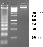
Apoptotic DNA laddering visualized in agarose gel. DNA from cells treated with an apoptotic inducing substance (left). A 1 kb marker (middle). Untreated cell DNA (right)

Gel electrophoresis is a standard technique that is frequently used to visualize DNA fragmentation (forming a ladder-like image on the gel), which is a characteristic of apoptosis and is associated with nuclear condensation (which characterizes pyknosis). Therefore, when referring to apoptosis, this technique is known as DNA laddering. Gel electrophoresis is also used to visualize the random DNA fragmentation of necrosis, which forms a smear on the gel.

Assays to detect DNA fragmentation or condensation

Various assays of DNA fragmentation or condensation include the APO single-stranded DNA (ssDNA) assay which detects damaged DNA of cells undergoing apoptosis or necrosis, TUNEL assay which is used to locally find DNA strand breaks (DSBs), and ISEL.[8]

ISEL (in-situ labeling technique) is a labeling/tagging technique of apoptotic or necrotic cells.[8] ISEL specifically targets unfragmented DNA that has condensed into a nucleosome structure.[8]

The APO ssDNA assay detects apoptotic cells by using an antibody that specifically binds to the ssDNA, which is accumulated during apoptosis as a result of DNA fragmentation.[8] Therefore, the presence of ssDNA is an indicator of DNA damage in the apoptotic cell. For the assay process, cells are fixed (with e.g., formamide), and these cells then undergo incubation (at a predetermined temperature), which subjects the DNA to thermal denaturation and exposes the ssDNA.[8] Next, the cells are incubated with an ssDNA-specific antibody along with a fluorescently labeled secondary antibody.[8] The fluorescence amounts as a measure of apoptosis which can then be quantified using flow cytometry.

The TUNEL assay, otherwise known as the terminal deoxynucleotidyl transferase dUTP nick-end labeling assay, is a technique that measures DNA damage and breakage during apoptosis.[8] During apoptosis, DNA fragmentation exposes numerous 3’OH ends, that are labeled with modified deoxy-uridine triphosphate (dUTP) by the TUNEL reaction.[8] Then, this modified dUTP can be identified with specific fluorescent antibodies which can identify modified nucleotides or by using tagged nucleotides themselves.[8] Flow cytometry can then be used to quantify fluorescence intensity, and thus provide a measure of apoptosis.

Detection methods of caspase activity

As mentioned above, caspase proteins, which are protease enzymes, promote DNA condensation and fragmentation via the caspase (or proteolytic) cascade. These caspase proteins include, for example, caspase 9, caspase 6, caspase 7, and caspase 3. The caspase cascade directly activates caspase-activated DNase (CAD) which initiates DNA fragmentation into smaller pieces resulting in chromatin condensation. The biochemical techniques used to detect caspase activity include ELISA and fluorometric and colorimetric assays.[8]

See also

References

  1. ^ Kumar V, Abbas A, Nelson F, Mitchell R (2007). Robbins Basic Pathology (8th ed.). pp. 6, 9–10 (table 1-1).
  2. ^ Kroemer G, Galluzzi L, Vandenabeele P, Abrams J, Alnemri ES, Baehrecke EH, et al. (January 2009). "Classification of cell death: recommendations of the Nomenclature Committee on Cell Death 2009". Cell Death and Differentiation. 16 (1): 3–11. doi:10.1038/cdd.2008.150. PMC 2744427. PMID 18846107.
  3. ^ Burgoyne, L. A. (1999-04-10). "The Mechanisms of Pyknosis: Hypercondensation and Death". Experimental Cell Research. 248 (1): 214–222. doi:10.1006/excr.1999.4406. ISSN 0014-4827. PMID 10094828.
  4. ^ a b c d e f g Liu, Lei; Gong, Fangyan; Jiang, Fang (2023-01-01), Boosani, Chandra S.; Goswami, Ritobrata (eds.), "Chapter 4 - Epigenetic regulation of necrosis and pyknosis", Epigenetics in Organ Specific Disorders, Translational Epigenetics, vol. 34, Academic Press, pp. 51–62, doi:10.1016/b978-0-12-823931-5.00024-4, ISBN 978-0-12-823931-5, retrieved 2024-12-05
  5. ^ a b Valenciano, Amy C.; Cowell, Rick L.; Rizzi, Theresa E.; Tyler, Ronald D. (2014-01-01), Valenciano, Amy C.; Cowell, Rick L.; Rizzi, Theresa E.; Tyler, Ronald D. (eds.), "Section 3 - White Blood Cells", Atlas of Canine and Feline Peripheral Blood Smears, Mosby, pp. 111–213.e2, doi:10.1016/b978-0-323-04468-4.00003-3, ISBN 978-0-323-04468-4, retrieved 2024-12-05
  6. ^ a b c d Hou, Lin; Liu, Kai; Li, Yuhong; Ma, Shuang; Ji, Xunming; Liu, Lei (2016-08-15). "Necrotic pyknosis is a morphologically and biochemically distinct event from apoptotic pyknosis". Journal of Cell Science. 129 (16): 3084–3090. doi:10.1242/jcs.184374. ISSN 0021-9533. PMID 27358477.
  7. ^ a b Ohara, Nobumasa; Uemura, Yasuyuki; Mezaki, Naomi; Kimura, Keita; Kaneko, Masanori; Kuwano, Hirohiko; Ebe, Katsuya; Fujita, Toshio; Komeyama, Takeshi; Usuda, Hiroyuki; Yamazaki, Yuto; Maekawa, Takashi; Sasano, Hironobu; Kaneko, Kenzo; Kamoi, Kyuzi (December 2016). "Histopathological analysis of spontaneous large necrosis of adrenal pheochromocytoma manifested as acute attacks of alternating hypertension and hypotension: a case report". Journal of Medical Case Reports. 10 (1). doi:10.1186/s13256-016-1068-3. ISSN 1752-1947. PMC 5059976. PMID 27729064.
  8. ^ a b c d e f g h i j Kari, Sana; Subramanian, Kumar; Altomonte, Ilenia Agata; Murugesan, Akshaya; Yli-Harja, Olli; Kandhavelu, Meenakshisundaram (2022-08-01). "Programmed cell death detection methods: a systematic review and a categorical comparison". Apoptosis. 27 (7): 482–508. doi:10.1007/s10495-022-01735-y. ISSN 1573-675X. PMC 9308588. PMID 35713779.

Read other articles:

James MasonMajalah Camera, 1922LahirJames Pier Mason(1889-02-03)3 Februari 1889Paris, PrancisMeninggal7 November 1959(1959-11-07) (umur 70)Hollywood, California, Amerika SerikatPekerjaanPemeranTahun aktif1914–1952 James Pier Mason (3 Februari 1889 – 7 November 1959) adalah seorang pemeran Amerika Serikat.[1] Ia tampil dalam 173 film antara 1914 dan 1952. Filmografi pilihan Film Tahun The Good Bad-Man 1916 On the Level 1917 Nan of Music Mountain 1917 Headin...


Koordinat: 5°03′43″S 119°31′57″E / 5.061868°S 119.5325167°E / -5.061868; 119.5325167 HasanuddinKelurahanKantor Kelurahan Hasanuddin di Jl. Bandara Lama No. 36Negara IndonesiaProvinsiSulawesi SelatanKabupatenMarosKecamatanMandaiKodepos90552[1]Kode Kemendagri73.09.01.1001 Kode BPS7308010011 Luas4,16 km² tahun 2017Jumlah penduduk9.049 jiwa tahun 2017Kepadatan2.175,24 jiwa/km² tahun 2017Jumlah RT32Jumlah RW6 Untuk pengertian lain, lihat Hasanuddi...


Embryonic precursor structures in vertebrates Pharyngeal archSchematic of developing human fetus with first, second and third arches labelledDetailsCarnegie stage10IdentifiersLatinarcus pharyngeiMeSHD001934TEarch_by_E5.4.2.0.0.0.2 E5.4.2.0.0.0.2 Anatomical terminology[edit on Wikidata] Floor of the pharynx of human embryo at about 26 days oldScheme of the pharyngeal archesScheme of the pharyngeal archesI–IV: pharyngeal arches1–4: pharyngeal pouches (inside) and/or pharyngeal grooves (...

Ukrainian public exhibition of captured Russian military equipment Destroyed Russian tanks in Mykhailivs'ka Square. The destroyed Russian military equipment exhibition is an open air exhibition on Mykhailivska Square in Kyiv.[1] It was opened on 21 May 2022, and features Russian military equipment that was captured and destroyed during the 2022 Russian invasion of Ukraine.[2][3][4] Exhibition on Mykhailivska Square According to the official website of the Presi...


Архангельский собор. Перспектива торцов надгробий царя Алексея Михайловича (1629—1676), царевича Алексея Алексеевича (1654—1670), царя Михаила Федоровича (1596—1645), царевичей-младенцев Василия и Ивана Михайловичей. Фотография К. А. Фишера. 1905 г. Из коллекций Музея архитект...


Spain Template‑class Spain portalThis template is within the scope of WikiProject Spain, a collaborative effort to improve the coverage of Spain on Wikipedia. If you would like to participate, please visit the project page, where you can join the discussion and see a list of open tasks.SpainWikipedia:WikiProject SpainTemplate:WikiProject SpainSpain articlesTemplateThis template does not require a rating on Wikipedia's content assessment scale. Trains: Rapid transit Template‑class Trains ...

Vilayet di ErzurumVilayet di Erzurum - LocalizzazioneIl vilayet di Erzurum nell'anno 1900 Dati amministrativiNome completoVilâyet-i Erzurum Nome ufficialeولايت ارضروم Lingue ufficialiturco ottomano Lingue parlateturco, curdo, armeno CapitaleErzurum Dipendente daImpero ottomano PoliticaForma di StatoVilayet Forma di governoVilayet elettivo dell'Impero ottomano Capo di StatoSultani ottomani Nascita1875 Fine1923 Territorio e popolazioneBacino geograficoTurchia Massima estensione76.70...


Artikel ini sebatang kara, artinya tidak ada artikel lain yang memiliki pranala balik ke halaman ini.Bantulah menambah pranala ke artikel ini dari artikel yang berhubungan atau coba peralatan pencari pranala.Tag ini diberikan pada Februari 2023. RedLink CommunicationsJenisPenyedia layanan internetDidirikan2008; 16 tahun lalu (2008)PendiriShane Thu Aung, Min Swe Hlaing, Thein Than ToeKantorpusatYangon, MyanmarWilayah operasiSeluruh negeriJasaAkses internet kecepatan tinggi kabel dan nirka...


Questa voce o sezione sull'argomento centri abitati della Campania non cita le fonti necessarie o quelle presenti sono insufficienti. Puoi migliorare questa voce aggiungendo citazioni da fonti attendibili secondo le linee guida sull'uso delle fonti. Conca della Campaniacomune Conca della Campania – Veduta LocalizzazioneStato Italia Regione Campania Provincia Caserta AmministrazioneSindacoDavid Simone (Risolleviamo Conca) dal 27-5-2019 TerritorioCoordinate41°2...

この記事は検証可能な参考文献や出典が全く示されていないか、不十分です。出典を追加して記事の信頼性向上にご協力ください。(このテンプレートの使い方)出典検索?: コルク – ニュース · 書籍 · スカラー · CiNii · J-STAGE · NDL · dlib.jp · ジャパンサーチ · TWL(2017年4月) コルクを打ち抜いて作った瓶の栓 コルク(木栓、�...


此條目可参照英語維基百科相應條目来扩充。 (2021年5月6日)若您熟悉来源语言和主题,请协助参考外语维基百科扩充条目。请勿直接提交机械翻译,也不要翻译不可靠、低品质内容。依版权协议,译文需在编辑摘要注明来源,或于讨论页顶部标记{{Translated page}}标签。 约翰斯顿环礁Kalama Atoll 美國本土外小島嶼 Johnston Atoll 旗幟颂歌:《星條旗》The Star-Spangled Banner約翰斯頓環礁�...


Oltrechiusa(IT) Oltrechiusa(LLD) Oltreciusa Stati Italia Regioni Veneto ( Belluno) TerritorioVal Boite, dalla chiusa di Venas a Dogana Vecchia CapoluogoSan Vito di Cadore Abitanti3 622 (31-08-2021) Lingueitaliano (ufficiale), ladino cadorino Il Pelmo da San Vito di Cadore L'Oltrechiusa (Oltreciusa in dialetto cadorino) è il territorio della valle del Boite posto oltre la chiusa di Venas e comprendente i comuni di Vodo, Borca e San Vito di Cadore. La chiusa era un...

Cet article est une ébauche concernant un aéroport chinois. Vous pouvez partager vos connaissances en l’améliorant (comment ?) selon les recommandations des projets correspondants. Aéroport d'Ordos Ejin Horo鄂尔多斯伊金霍洛机场È'ěrduōsī Yījīn Huòluò Jīchǎng Terminal de l'aéroport. Localisation Pays Chine Ville Ordos Coordonnées 39° 29′ 38″ nord, 109° 51′ 44″ est Informations aéronautiques Code IATA DSN Code OACI ZBDS T...


Bahasa SasakBPS: 0164 4 ᬪᬵᬲᬵᬲᬓ᭄ᬱᬓ᭄ Dituturkan diIndonesiaWilayah Nusa Tenggara Barat Pulau Lombok EtnisSasakPenutur2,7 juta[1] (2010) Rumpun bahasa Austronesia Melayu-Polinesia Melayu-Sumbawa (?) Bali-Sasak-Sumbawa Sasak-Sumbawa Sasak Untuk kontributor: Sedang dilakukan otomatisasi klasifikasi bahasa secara berkala. Silakan sampaikan saran, pendapat, maupun perbaikan pada halaman pembicaraan templat maupun pembicaraan ProyekWiki Sistem penulisanSasak...


الرابطة الوطنية لكرة القدم المحترفة الاسم المختصر LNFP الرياضة كرة قدم أسس عام 1994 الرئيس محمد العربي المقر تونس العاصمة الانتسابات الجامعة التونسية لكرة القدم النوادي 46 الموقع الرسمي www.ftf.org.tn تعديل مصدري - تعديل   الرابطة الوطنية لكرة القدم المحترفة (بالفرنسية: Ligue nationale de...

  لمعانٍ أخرى، طالع أبو سيف (توضيح). اضغط هنا للاطلاع على كيفية قراءة التصنيف أبو سيفالعصر: الفترة الضحوية–الآن قك ك أ س د ف بر ث ج ط ب ن [1] حالة الحفظ   أنواع قريبة من خطر الانقراض [2] المرتبة التصنيفية نوع  التصنيف العلمي النطاق: حقيقيات النوى المملكة: الحيوا...


Titan IIIC adalah booster ruang angkasa yang digunakan oleh Angkatan Udara Amerika Serikat. Ini direncanakan untuk digunakan sebagai kendaraan peluncuran di dibatalkan Dyna-Soar dan program Manned Orbiting Laboratory. Titan III juga digunakan untuk meluncurkan beberapa satelit selama misi tunggal. Ini diluncurkan secara eksklusif dari Cape Canaveral, sementara saudaranya Titan IIID diluncurkan hanya dari Vandenberg AFB. Mayoritas payload IIIC itu diklasifikasikan satelit DoD, terutama pengin...


Artikel ini sebatang kara, artinya tidak ada artikel lain yang memiliki pranala balik ke halaman ini.Bantulah menambah pranala ke artikel ini dari artikel yang berhubungan atau coba peralatan pencari pranala.Tag ini diberikan pada November 2022. Cha Cha for TwinsSutradaraYang Yi-chienJim WangPemeranHuang PeijiaTanggal rilis 15 Juni 2012 (2012-06-15) (Taiwan) 10 Januari 2013 (2013-01-10) (Tiongkok) NegaraTaiwanBahasaMandarin Cha Cha for Twins (Hanzi: 寶米恰恰; Bao mi ...

Architectural, interior, product, and graphic design of the mid-20th century This section needs additional citations for verification. Please help improve this article by adding citations to reliable sources in this section. Unsourced material may be challenged and removed. (March 2012) (Learn how and when to remove this message) Mid-century modern architectureTract house in Tujunga, California, featuring open-beamed ceilings, c. 1960Years active1945–1970LocationUnited StatesInfluencesI...


Mogami-Klasse Die Mogami im Jahr 1935 bei einer Probefahrt. Die Mogami im Jahr 1935 bei einer Probefahrt. Schiffsdaten Land Japan Japan Schiffsart Leichter Kreuzer(1934–1939)Schwerer Kreuzer(1939–1944) Bauzeitraum 1931 bis 1937 Stapellauf des Typschiffes 14. März 1934 Gebaute Einheiten 4 Dienstzeit 1935 bis 1944 Schiffsmaße und Besatzung Länge 200,60 m (Lüa)197 m (KWL)189 m (Lpp) Breite 18,00 m Tiefgang (max.) 5,50 m Verdrängung 8636 t   Besatzung 850 Mann Maschinenan...