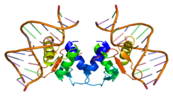
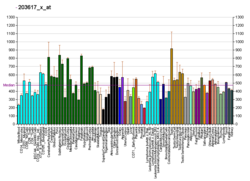
ELK1
|
Read other articles:

This article needs additional citations for verification. Please help improve this article by adding citations to reliable sources. Unsourced material may be challenged and removed.Find sources: List of NBA All-Star Game records – news · newspapers · books · scholar · JSTOR (June 2022) (Learn how and when to remove this template message) This article lists all-time leading figures achieved in the NBA All-Star Game in every major statistical category r...

1996 Japanese animated film by Rintaro XTheatrical release poster, featuring Kamui Shiro (left) and Fuma Monou (right)Directed byRintaroScreenplay byRintaroNanase OhkawaBased onXby ClampProduced byKazuhiko IkeguchiKazuo YokoyamaMasao MaruyamaStarringTomokazu SekiKen NaritaCinematographyHitoshi YamaguchiEdited byHarutoshi OgataSatoshi TerauchiYukiko ItoMusic byYasuaki ShimizuProductioncompanyMadhouseDistributed byToei CompanyRelease dates August 3, 1996 (1996-08-03) (Japan) ...

2020 benefit livestream event Together at HomeAlso known asOne World: Together at HomeCreated byGlobal CitizenWritten byGlobal CitizenPresented byJimmy FallonJimmy KimmelStephen ColbertOriginal languageEnglishProductionExecutive producersAudrey Morrissey[1]Hugh Evans[2]Lee RolontzProduction locationVirtualRunning time120 minutesProduction companyGlobal CitizenOriginal releaseNetworkSyndicationReleaseApril 18, 2020 (2020-04-18)RelatedGlobal Goal: Unite for Our Fu...

Синелобый амазон Научная классификация Домен:ЭукариотыЦарство:ЖивотныеПодцарство:ЭуметазоиБез ранга:Двусторонне-симметричныеБез ранга:ВторичноротыеТип:ХордовыеПодтип:ПозвоночныеИнфратип:ЧелюстноротыеНадкласс:ЧетвероногиеКлада:АмниотыКлада:ЗавропсидыКласс:Пт�...

Autoloading rifle Zastava M92 Zastava M92TypeCarbinePlace of originYugoslavia/SerbiaService historyIn service1992–presentWarsYugoslav WarsKivu conflict[1]Libyan Civil WarBanjska attackProduction historyManufacturerZastava ArmsProducedsince 1992SpecificationsMass3.57 kg (7.87 lb)Length795 mm (31.30 in) stock extended 550 mm (21.65 in) stock foldedBarrel length254 mm (10.0 in)Cartridge7.62×39mm[2]ActionGas-operated, ...

Запрос «Пугачёва» перенаправляется сюда; см. также другие значения. Алла Пугачёва На фестивале «Славянский базар в Витебске», 2016 год Основная информация Полное имя Алла Борисовна Пугачёва Дата рождения 15 апреля 1949(1949-04-15) (75 лет) Место рождения Москва, СССР[1]...

Former and Westernmost gate in London Wall For the surname, see Ludgate (surname). LudgateAn old illustration of the gate c. 1650General informationTown or cityLondonCountryEnglandCoordinates51°30′50.3″N 0°06′08.2″W / 51.513972°N 0.102278°W / 51.513972; -0.102278 Ludgate was the westernmost gate in London Wall. Of Roman origin, it was rebuilt several times and finally demolished in 1760. The name survives in Ludgate Hill, an eastward continuation of F...

Swedish politician (born 1966) Hans EklindMember of the RiksdagIncumbentAssumed office 24 September 2018ConstituencyÖrebro County Personal detailsBorn1966 (age 57–58)Political partyChristian Democrats Hans Eklind (born 1966)[1] is a Swedish politician. Since September 2018,[update] he serves as Member of the Riksdag from the Christian Democrats representing the constituency of Örebro County.[2] He was also elected as Member of the Riksdag in Septe...

This article is about the Northern Irish politician. For other people named James McCusker, see James McCusker. British politician Harold McCuskerMPMember of Parliament for Upper BannIn office9 June 1983 – 12 February 1990Preceded bySeat createdSucceeded byDavid TrimbleMember of Parliament for ArmaghIn office28 February 1974 – 13 May 1983Preceded byJohn MaginnisSucceeded bySeat abolished Personal detailsBornJames Harold McCusker(1940-02-07)7 February 1940Lurgan, Northern...

Danish politician Brian MikkelsenCEO of the Danish Chamber of CommerceIncumbentAssumed office 21 June 2018Minister of Industry, Business and Financial AffairsIn office28 November 2016 – 20 June 2018Prime MinisterLars Løkke RasmussenPreceded byTroels Lund PoulsenSucceeded byRasmus JarlovMinister of Economic and Business AffairsIn office23 February 2010 – 3 October 2011Prime MinisterLars Løkke RasmussenPreceded byLene EspersenSucceeded byOle SohnMinister of JusticeIn...

Verschiedene RFID-Transponder Universelles RFID-Handlesegerät für 125 kHz, 134 kHz und 13,56 MHz; optional Barcode Gerät für UHF Tags, 630 mW Leistung RFID-Bluetooth-Handlesegerät für 13,56 MHz, mit Ferritantenne zum Auslesen sehr kleiner Transponder aus Metall Mobiles Android-5.1-Gerät mit LF- und HF-RFID in einem Gerät RFID (englisch radio-frequency identification [ˈɹeɪdɪəʊ ˈfɹiːkwənsi aɪˌdɛntɪfɪˈkeɪʃn̩] „Identifizierung mit Hilfe elektromagn...

Domestic airport in Kullu, Himachal Pradesh, India Kullu–Manali AirportIATA: KUUICAO: VIBRSummaryAirport typePublicOwnerGovernment of IndiaOperatorAirports Authority of IndiaServesKullu, ManaliLocationBhuntar, Kullu, Himachal Pradesh, IndiaElevation AMSL1,089 m / 3,573 ftCoordinates31°52′36″N 77°09′16″E / 31.87667°N 77.15444°E / 31.87667; 77.15444MapKUULocation of airport in IndiaShow map of Himachal PradeshKUUKUU (India)Show map of IndiaRu...

2006年民主進步黨主席補選,正式名稱為民主進步黨第十一屆第二次主席補選,是民主進步黨自1998年第八屆黨主席選舉以來所舉辦的第四次黨員直選黨主席,因為民進黨在2005年中華民國地方公職人員選舉慘敗後,當時的黨主席蘇貞昌為了表示負責即宣佈辭去黨主席一職。 民主進步黨第十一屆第二次主席補選 ← 2005 2006年1月15日 2008 → 候选人 游錫堃 蔡同榮 ...

Pour les articles homonymes, voir Éclipse (homonymie). Si ce bandeau n'est plus pertinent, retirez-le. Cliquez ici pour en savoir plus. Cet article ne cite pas suffisamment ses sources (novembre 2020). Si vous disposez d'ouvrages ou d'articles de référence ou si vous connaissez des sites web de qualité traitant du thème abordé ici, merci de compléter l'article en donnant les références utiles à sa vérifiabilité et en les liant à la section « Notes et références ». ...

Menteri Kesehatan MalaysiaMenteri Kesihatan Malaysia منتري كصيحتن مليسياLambangPetahanaDzulkefly Ahmadsejak 12 Desember 2023Kementerian KesehatanGelarYang Berhormat Menteri(Yang Terhormat Menteri)Ditunjuk olehYang di-Pertuan Agong atas petunjuk Perdana Menteri MalaysiaDibentuk9 Agustus 1955; 68 tahun lalu (1955-08-09)Pejabat pertamaLeong Yew KohSitus webwww.moh.gov.my Berikut adalah daftar orang yang pernah menjabat sebagai Menteri Kesehatan (bahasa Melayu: Ment...

Kwik Djoen Eng pada 1921 Kwik Djoen Eng (Hanzi: 郭春秧) atau Kwok Chun Yeung (1860-1935) adalah seorang pengusaha asal Taiwan. Ia merupakan salah satu pendiri firma dagang terkenal Kwik Hoo Tong (KHT) di Solo, Hindia Belanda (sekarang Indonesia).[1] Ia dijuluki sebagai Raja Gula Jawa namun memiliki kepentingan komersial di Hong Kong.[2] Ia juga merupakan bekas pemilik Istana Djoen Eng yang berada di Salatiga namun kemudian disita oleh Javaasche Bank pada 1930 saat seb...

Relazioni tra India e Italia Mappa che indica l'ubicazione di India e Italia India Italia Le relazioni bilaterali tra India e Italia fanno riferimento ai rapporti diplomatici ed economici tra la Repubblica d'India e la Repubblica Italiana. L'India ha un'ambasciata a Roma e un Consolato Generale a Milano. L'Italia ha un'ambasciata a Nuova Delhi e due Consolati Generali a Mumbai e Calcutta. Indice 1 Storia delle relazioni...

西班牙共產黨Partido Comunista de España西班牙共產黨标志主席何塞·路易斯·森特利亚总书记恩里克·圣地亚哥名誉主席多洛雷斯·伊巴露丽[1]成立1921年11月14日 (1921-11-14)合并自西班牙的共產黨西班牙共產主义工人黨总部马德里党报《工人世界》《我们的旗帜》(杂志)智庫马克思主义研究基金会青年组织西班牙共产主义青年联盟妇女组织妇女民主运动(1965年—1976年,20...

American politician (1865–1951) For the housing project in Washington, D.C, see Arthur Capper/Carrollsburg. Arthur CapperCapper in 1905United States Senatorfrom KansasIn officeMarch 4, 1919 – January 3, 1949Preceded byWilliam ThompsonSucceeded byAndrew SchoeppelChair of the National Governors AssociationIn officeDecember 14, 1916 – September 16, 1924Preceded byWilliam SprySucceeded byEmerson Harrington20th Governor of KansasIn officeJanuary 11, 1915 – Januar...

Indoor arena at the University of Connecticut Gampel PavilionThe Basketball Capital of the WorldLocation2095 Hillside RoadStorrs-Mansfield, CT, United States 06269Coordinates41°48′19.05″N 72°15′15.10″W / 41.8052917°N 72.2541944°W / 41.8052917; -72.2541944OwnerUniversity of ConnecticutOperatorUniversity of ConnecticutCapacity2023–present: 10,2992002–2023: 10,1671996–2002: 10,0271990–1996: 8,241[1]Surface171,000 sq ft (15,900 m2...


