Neural plate

Neural plate
Neural crest
Details
Carnegie stage9
Days19
PrecursorEctoderm
Gives rise toNeural folds
SystemCentral nervous system
Identifiers
Latinlamina neuralis
MeSHD054258
TEplate_by_E5.13.1.0.1.0.1 E5.13.1.0.1.0.1
Anatomical terminology

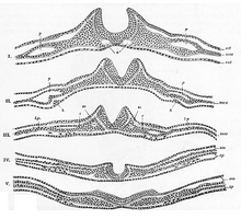
In embryology, the neural plate is a key developmental structure that serves as the basis for the nervous system. Cranial to the primitive node of the embryonic primitive streak, ectodermal tissue thickens and flattens to become the neural plate. The region anterior to the primitive node can be generally referred to as the neural plate. Cells take on a columnar appearance in the process as they continue to lengthen and narrow. The ends of the neural plate, known as the neural folds, push the ends of the plate up and together, folding into the neural tube, a structure critical to brain and spinal cord development. This process as a whole is termed primary neurulation.[1]

Signaling proteins are also important in neural plate development, and aid in differentiating the tissue destined to become the neural plate. Examples of such proteins include bone morphogenetic proteins and cadherins. Expression of these proteins is essential to neural plate folding and subsequent neural tube formation.

Involvement in primary neurulation

Neurulation

Generally divided into four, the process of primary neurulation involves the neural plate in the first three steps. The formation and folding of the neural plate is the first step in primary neurulation. This is followed by the refinement and growth of neural plate cells. The third step of primary neurulation does not involve the neural plate per se, but rather the edges of the neural plate, which come together, turning the plate into the start of the neural tube. With the neural plate having folded into a tube, the neural folds come together to complete the fusion of the neural tube. This process is illustrated in the figure to the right, where the neural plate is shown in purple. The lime green marks the edges of the neural plate, which become the neural folds, involved in the folding of the plate to create the neural tube. The figure demonstrates the development of the neural plate into the neural tube, which is where the neural crest cells are derived from as well.[1]

In primary neurulation, the layer of ectoderm divides into three sets of cells: the neural tube (future brain and spinal cord), epidermis (skin), and neural crest cells (connects epidermis and neural tube and will migrate to make neurons, glia, and skin cell pigmentation).[1]

Development

During the stage of neural plate formation the embryo consists of three cell layers: the ectoderm that eventually forms the skin and neural tissues, the mesoderm that forms muscle and bone, and the endoderm that will form the cells lining the digestive and respiratory tracts. The progenitor cells that make up the precursors to neural tissues in the neural plate are called neuroepithelial cells.[citation needed]

Stretched over the notochord, the ectodermal cells on the dorsal portion of the embryo are ultimately the ones that form the neural plate. Approximately half of those cells will be induced to remain ectoderm, while the other half will form the neural plate.[2][3]

There are four stages of neural plate and neural tube formation: formation, bending, convergence, and closure. The formation of the neural plate starts when dorsal mesoderm signals ectodermal cells above it to lengthen into columnar neural plate cells.[4] This different shape distinguishes the cells of the presumptive neural plate from other pre-epidermal cells. If the neural plate is separated by itself, it will still develop to make a thinner plate but will not form a neural tube. If the region containing presumptive epidermis and neural plate tissue is isolated, small neural folds will form. Elongation that occurs throughout the formation of the neural plate and closure of the neural tube is vital; the closing areas of the neural tube are seen to have very increased elongation activity in the midline compared to already closed areas when the plate was beginning to shape itself into a tube.[5]

Bending and Convergence of the Neural Plate

The bending of the neural plate involves the formation of hinges, where the neural plate is connected to surrounding tissues. The midline of the neural plate is referred to the median hinge point (MHP). Cells in this area, known as medial hinge point cells because of their involvement with this structure, are stabilized and connected to the notochord. They are derived from the area of the neural plate anterior to primitive knot. The notochord will begin the shape changes in MHP cells. These cells will decrease in height and become wedge-shaped. Another type of hinge point occurs dorsal-laterally, referred to as dorsal-lateral hinge point (DLHP). These regions furrow and change shape in the same way as MHP cells do before connecting together to form the neural tube. It was seen in an experiment that without the notochord, the MHP characteristics did not develop correctly, so the neural plate and neural tube formation did not happen properly.[6] The communication between the neural plate and the notochord is important for the future induction and formation of the neural tube.

Closure of the neural tube is completed when the neural folds are brought together, adhering to each other. While the cells that remain as the neural tube form the brain and spinal cord, the other cells that were part of the neural plate migrate away from the tube as neural crest cells. After an epithelial–mesenchymal transition, these cells form the autonomic nervous system and certain cells of the peripheral nervous system.[7]

Cell signaling and essential proteins

Critical to the proper folding and function of the neural plate is N-cadherin, a type of cadherin protein associated with the nervous system. N-cadherin is critical to holding neural plate cells together. Additionally, cells destined to become neural plate cells express nerve cell adhesion molecule (NCAM) to further neural plate cohesion. Another cadherin, E-cadherin, is expressed by ectodermal cells in the process of neural plate development.[1]

3-D structural model of BMP-4

Bone morphogenetic protein 4, or BMP4, is a transforming growth factor that causes the cells of the ectoderm to differentiate into skin cells. Without BMP4 the ectoderm cells would develop into neural cells. Axial mesoderm cells under the ectoderm secrete inhibitory signals called chordin, noggin and follistatin. These inhibitory signals prevent the action of BMP4, which would normally make the cells ectoderm; as a result, the overlying cells take their normal course and develop into neural cells. The cells in the ectoderm that circumscribe these neural cells do not receive the BMP4 inhibitor signals and as a result BMP4 induces these cells to develop into skin cells.[8]

Neural plate border specifiers are induced as a set of transcription factors. Distalless-5, PAX3 and PAX7 prevent the border region from becoming either neural plate or epidermis.[1] These induce a second set of transcription factors called neural crest specifiers, which cause cells to become neural crest cells.

In a newly formed neural plate, PAX3 mRNA, MSX1 mRNA, and MSX1/MSX2 proteins are expressed mediolaterally.[9] When the neural plate begins to fold, rostral areas of the neural plate do not express Pax3 and MSX proteins. Areas caudal to neural tube closure have PAX3 and MSX expression restricted to lateral regions of the neural folds.[9] These fluctuations in mRNA and protein expression allude to how they play a role in differentiation of neural plate cells.

Low pSMAD 1, 5, 8 levels allow a greater mobility at the median hinge point than in lateral neural plate cells.[10] This flexibility allows for the pivoting and hinging that allows the buckling and lifting of the neural plate when formatting the neural tube. The neural plate has to be rigid enough for morphogenic movements to occur while being flexible enough to undergo shape and position changes for the transformation to the neural tube.

Other animals

The neural tube closes differently in various species, the distinctions between humans and chickens being some of the most studied. In humans, the neural tube fuses together from a central region of the embryo and moves outwards. In chickens, neural tube closure begins at the future midbrain region and it closes in both directions.[1] In birds and mammals, the closure does not occur at the same time.

In newt and general amphibian embryos, cell division is not a driving role in morphogenesis. Newt embryo cells are much larger and exhibit egg pigmentation to distinguish cells from each other. The newt neural plate doubles in length, decreases in apical width, and increases in thickness.[5] The plate edges rise dorsally and fold toward the midline to form the neural tube. The apical surface area decreases.

In chicken embryos, while the neural plate increases in length and decreases in apical width, the thickness of the plate does not change drastically. As the neural plate progresses through the Hamburger-Hamilton stages, the plate thickens until about HH6-7, when the neural plate begins to fold into tube form. The apical surface area increases during neurulation, unlike amphibian embryos.[5] In mouse embryos, there is a large convex-shaped curve to each side of the middle of the plate. This curve has to be reversed as the plate rolls together to form the neural tube.[5]

Research

Research on the neural plate began in earnest by looking into the determination of the ectoderm and its commitment to the neuronal path. With the development of research and laboratory techniques there have been major advances in the study of neurulation and the development and role of the neural plate in a growing embryo. The use of such techniques vary with the stage of development and overall research goals, but include such methods as cell labeling and grafting.[11]

Cell labelling

The process of in situ hybridization (ISH) follows the labeling of a DNA or RNA sequence to serve as an antisense mRNA probe, complementary to a sequence of mRNA within the embryo. Labeling with a fluorescent dye or radioactive tag allows for the visualization of the probe and their location within the embryo. This technique is useful as it reveals specific areas of gene expression in a tissue as well as throughout an entire embryo through whole-mount in situ hybridization.[12] This technique is often used in determination of gene expression necessary for the proper development of the embryo. Marking certain genes in a developing embryo allows for the determination of the exact time and place in which the gene is activated, offering information in the role of the particular gene in development.

Similar to the process of in situ hybridization, immunofluorescence (IF) also allows for the determination of particular cell element's roles in development. In contrast to in situ hybridization however, immunofluorescence uses a fluorophore attached to an antibody with biomolecule target, such as proteins, rather than DNA and RNA sequences. The allows for the visualization of biomolecule elements of the cell. In the study of embryogenesis immunofluorescence may be used for purposes similar to hybridization, for the tracking of proteins that are involved in the development of the embryo and their specific time and place of production and use.[13] Current research has expanded on the immunofluorescence technique to combined it with the methods of in situ hybridization, either fluorescent or radioactive. This combination is believed to increase specificity and take away for the limitations of each individual technique. For example, this method with enhance counterstaining in a tissue and multiple protein labeling.[12]

Cell grafting

Cell grafting in the early stages of embryo development has provided crucial information on cell fates and the processes of determination. Grafting at specific stages of neurulation has advanced research on the signaling necessary for the proper development of the neural plate and other structures. The grafting of the ectoderm and neural structures is very specialized and delicate procedure, requiring the removal and marking of a desired group of cells, followed by their transplantation, for example, into a new area of the embryo.[14]

Grafting experiments done in Xenopus and chicken embryos show the neural plate's capability to induce other regions of cells, including the pre-placodal region, a group of ectodermal cells essential to the function of sensory organs.[15]

References

Public domain This article incorporates text in the public domain from the 20th edition of Gray's Anatomy (1918)

  1. ^ a b c d e f Gilbert, Scott F. (2010). Developmental biology (9th. ed.). Sunderland, Mass.: Sinauer Associates. pp. 333–338. ISBN 978-0878933846.
  2. ^ Browder, Leon (1980). Developmental Biology. Philadelphia: Saunders College. p. 457. ISBN 0-03-056748-3.
  3. ^ Human Embryology, Module 7, Section 7.2, http://www.embryology.ch/anglais/hdisqueembry/triderm10.html Archived 2013-01-16 at the Wayback Machine.
  4. ^ Keller, Ray; Shih, John; Sater, Amy K (1 March 1992). "Planar induction of convergence and extension of the neural plate by the organizer Xenopus". Developmental Dynamics. 193 (3): 218–234. doi:10.1002/aja.1001930303. PMID 1600241. S2CID 39722561.
  5. ^ a b c d Jacobson, Antone G. (1991). "Experimental Analyses of the Shaping of the Neural Plate and Tube". American Zoologist. 31 (4): 628–643. doi:10.1093/icb/31.4.628. JSTOR 3883562.
  6. ^ Smith, Jodi L.; Schoenwolf, Gary C. (1 April 1989). "Notochordal induction of cell wedging in the chicken neural plate and its role in neural tube formation". Journal of Experimental Zoology. 250 (1): 49–62. doi:10.1002/jez.1402500107. PMID 2723610.
  7. ^ Wolpert, Lewis (1998). Principles of Development. London: Current Biology. p. 345. ISBN 0-19-850263-X.
  8. ^ Wilson, PA; Lagna, G; Suzuki, A; Hemmati-Brivanlou, A (Aug 1997). "Concentration-dependent patterning of the Xenopus ectoderm by BMP4 and its signal transducer Smad1". Development. 124 (16): 3177–84. doi:10.1242/dev.124.16.3177. PMID 9272958.
  9. ^ a b Liem, Karel F; Tremml, Gabi; Roelink, Henk; Jessell, Thomas M (1 September 1995). "Dorsal differentiation of neural plate cells induced by BMP-mediated signals from epidermal ectoderm". Cell. 82 (6): 969–979. doi:10.1016/0092-8674(95)90276-7. PMID 7553857. S2CID 17106597.
  10. ^ Eom, Dae S; Amarnath, Smita; Agarwala, Seema (20 December 2012). "Apicobasal Polarity and neural tube closure". Development, Growth & Differentiation. 55 (1): 164–172. doi:10.1111/dgd.12030. PMC 3540145. PMID 23277919.
  11. ^ de Vellis J, Carpenter E. General Development of the Nervous System. In: Siegel GJ, Agranoff BW, Albers RW, et al., editors. Basic Neurochemistry: Molecular, Cellular and Medical Aspects. 6th edition. Philadelphia: Lippincott-Raven; 1999. Available from: https://www.ncbi.nlm.nih.gov/books/NBK28253/
  12. ^ a b Pineau, Isabelle (2006). "A Novel Method for Multiple Labeling Combining In Situ Hybridization With Immunofluorescence". Journal of Histochemistry & Cytochemistry. 54 (11): 1303–1313. doi:10.1369/jhc.6a7022.2006. PMID 16899759.
  13. ^ Sadler, T.W. (1986). "A potential role for spectrin during neurulation". J. Embryol. 94 (1): 73–82. Retrieved 27 April 2013.
  14. ^ Tan, SS (1986). "Analysis of cranial neural crest cell migration and early fates in postimplantation rat chimaeras". J. Embryol. 98 (1): 21–58. PMID 3655649. Retrieved 27 April 2013.
  15. ^ Bailey, Andrew P.; Andrea Streit (2006). "Sensory Organs: Making and Breaking the Pre-Placodal Region". Current Topics in Developmental Biology. 72: 177. doi:10.1016/s0070-2153(05)72003-2. ISBN 9780121531720. PMID 16564335.

Read other articles:

Newcastle United Jets FCNama lengkapNewcastle United Jets FCJulukanJetsBerdiri2000StadionStadion EnergyAustralia, Newcastle(Kapasitas: 26,164)Ketua Con ConstantineManajer Gary van EgmondLigaA-League2018-197 Kostum kandang Kostum tandang Newcastle United Jets FC merupakan sebuah tim sepak bola Australia yang bermain di divisi utama A-League. Didirikan pada tahun 2000. Berbasis di Newcastle. Klub ini memainkan pertandingan kandangnya di Stadion EnergyAustralia yang berkapasitas 26.164 kursi. Se...


United States historic placeJohnson's Grocery StoreU.S. National Register of Historic Places Johnson's Grocery StoreLocation301 N. Picacho, Casa Grande, ArizonaCoordinates32°52′36″N 111°45′02″W / 32.87667°N 111.75056°W / 32.87667; -111.75056 (Johnson's Grocery Store)Arealess than one acreBuiltc.1907MPSCasa Grande MRANRHP reference No.85000885[1]Added to NRHPApril 16, 1985 Johnston's Grocery Store, at 301 N. Picacho in Casa Grande, ...


Abarema filamentosa Status konservasi Rentan  (IUCN 2.3)[1] Klasifikasi ilmiah Kerajaan: Plantae (tanpa takson): Angiospermae (tanpa takson): Eudikotil (tanpa takson): Rosidae Ordo: Fabales Famili: Fabaceae Genus: Abarema Spesies: A. filamentosa Nama binomial Abarema filamentosa(Bentham) Pittier Sinonim Feuilleea filamentosa (Benth.) Kuntze Pithecellobium filamentosum Benth. Pithecolobium filamentosum Benth. Sebuah tumbuhan Abarema filamentosa yang sedang berbunga di alam l...

2009 film directed by Mamoru Hosoda Summer WarsTheatrical release posterJapanese nameKanjiサマーウォーズTranscriptionsRevised HepburnSamā Wōzu Directed byMamoru HosodaScreenplay bySatoko OkuderaStory byMamoru HosodaProduced byNozomu TakahashiTakuya ItoTakafumi WatanabeYuichiro SaitoStarringRyunosuke KamikiNanami SakurabaMitsuki TanimuraSumiko FujiCinematographyYukihiro MatsumotoEdited byShigeru NishiyamaMusic byAkihiko MatsumotoProductioncompanyMadhouseDistributed byWarner Bros. Pict...


Poultry farm in Arcadia, Wisconsin Yarding poultry farm in Vernon County, Wisconsin Battery chickens Poultry farming is a part of the United States's agricultural economy. History This section is missing information about Native American uses and domestication of poultry. Please expand the section to include this information. Further details may exist on the talk page. (September 2022) The best in the world White Plymouth Rocks, 1910 In the United States, chickens were raised primarily on fa...


Découpage cantonal du département des Hautes-Alpes, avec en surimpression les arrondissements (en nuances de bleu) - Carte arrêtée au 1er janvier 2019. Le département des Hautes-Alpes compte 15 cantons depuis le redécoupage cantonal de 2014 (30 cantons auparavant). Histoire Découpage cantonal antérieur à 2015 Localisation géographique des chefs-lieux de canton des Hautes-Alpes Le département des Hautes-Alpes compte 30 cantons : 7 cantons dans l'arrondissement de Briançon ...

Gavirate komune di Italia Gavirate (it) Tempat Negara berdaulatItaliaRegion di ItaliaLombardyProvinsi di ItaliaProvinsi Varese NegaraItalia Ibu kotaGavirate PendudukTotal9.136  (2023 )Bahasa resmiItalia GeografiLuas wilayah12,01 km² [convert: unit tak dikenal]Ketinggian261 m Berbatasan denganBarasso Bardello con Malgesso e Bregano Besozzo Casciago Comerio Varese Biandronno Cocquio-Trevisago Cuvio SejarahSanto pelindungYohanes sang Penginjil Informasi tambahanKode pos21026 Zona wakt...


2016年美國總統選舉 ← 2012 2016年11月8日 2020 → 538個選舉人團席位獲勝需270票民意調查投票率55.7%[1][2] ▲ 0.8 %   获提名人 唐納·川普 希拉莉·克林頓 政党 共和黨 民主党 家鄉州 紐約州 紐約州 竞选搭档 迈克·彭斯 蒂姆·凱恩 选举人票 304[3][4][註 1] 227[5] 胜出州/省 30 + 緬-2 20 + DC 民選得票 62,984,828[6] 65,853,514[6]...


Ř

Ř i versal- och gemen form. Ř (gemenform: ř) är den latinska bokstaven R med det diakritiska tecknet hake över. Ř finns i det tjeckiska, schlesiska och översorbiska alfabetet. Tjeckiska På tjeckiska representerar Ř ljudet [r̝]. Detta ljud finns i mycket få andra språk och brukar vara det sista ljudet som behärskas av tjeckiska barn. En populär tjeckisk tungvrickare som innehåller bokstaven i fråga är: Tři sta třicet tři stříbrných stříkaček stříkalo přes tři sta ...

كأس تركيا 2017–18 تفاصيل الموسم كأس تركيا  النسخة 56  البلد تركيا  التاريخ بداية:22 أغسطس 2017  نهاية:10 مايو 2018  المنظم اتحاد تركيا لكرة القدم  البطل نادي بلدية آكهيسار سبور  مباريات ملعوبة 188   عدد المشاركين 159   كأس تركيا 2016–17  كأس تركيا 2018–19  تعديل مصد...


River in AfghanistanHarut RiverAdraskan RiverNative nameهاروت رود (Pashto)LocationCountryAfghanistanPhysical characteristicsSource  • locationSia koh mountains Mouth  • locationSistan LakeLength394 km (245 mi) [1]Basin sizeSistan Basin The Harut River or Adraskan River is a river of Afghanistan which belongs to the Sistan Basin.[2] The source of the river lies in the mountains to the southeast of Herat. The ...


Professional wrestling trios tag team championship WCWA World Tag Team ChampionshipChampionship beltDetailsPromotionWorld Class Championship Wrestling[1][2]World Class Wrestling Association[1][2]Date establishedDecember 25, 1982[1][2]Date retired1988[1][2]Other name(s) WCCW World Six-Man Tag Team Championship[1][2] NWA World Six-Man Tag Team Championship (Texas version)[1][2] StatisticsFirst champi...

Nationality law of Swaziland, Africa Swaziland Citizenship ActParliament of EswatiniEnacted byGovernment of EswatiniStatus: Current legislation Eswatini nationality law is regulated by the Constitution of Eswatini, as amended; the Swaziland Citizenship Act, and its revisions; and various international agreements to which the country is a signatory.[1][2] These laws determine who is, or is eligible to be, a national of Eswatini.[3] The legal means to acquire nation...


2021 film by Steven Spielberg West Side StoryTheatrical release posterDirected bySteven SpielbergScreenplay byTony KushnerBased onWest Side Storyby Jerome Robbins Leonard Bernstein Stephen Sondheim Arthur LaurentsProduced by Steven Spielberg Kristie Macosko Krieger Kevin McCollum Starring Ansel Elgort Ariana DeBose David Alvarez Mike Faist Rita Moreno Rachel Zegler CinematographyJanusz KamińskiEdited by Michael Kahn Sarah Broshar Music byLeonard BernsteinProductioncompanies Amblin Entertainm...


Artikel ini sebatang kara, artinya tidak ada artikel lain yang memiliki pranala balik ke halaman ini.Bantulah menambah pranala ke artikel ini dari artikel yang berhubungan atau coba peralatan pencari pranala.Tag ini diberikan pada Februari 2023. Artikel ini bukan mengenai Ganten, Kerjo, Karanganyar. Ganten景田 Negara Tiongkok Sumber {{{source}}} Jenis {{{type}}} TDS {{{tds}}} miligram per liter (mg/l) Situs web: www.ganten.com.cn Ganten (Hanzi: 景田; Pinyin: Jǐngtián; Jyutping&#...

  لمعانٍ أخرى، طالع سنترال سيتي (توضيح). هذه المقالة يتيمة إذ تصل إليها مقالات أخرى قليلة جدًا. فضلًا، ساعد بإضافة وصلة إليها في مقالات متعلقة بها. (سبتمبر 2014) سنترال سيتي   الإحداثيات 30°33′16″N 91°02′12″W / 30.554444444444°N 91.036666666667°W / 30.554444444444; -91.036666666667   تاريخ ا�...

 

Newspaper The Chronicle of Higher EducationSeptember 18, 2009 front page of The ChronicleTypeWeekly newspaper, websiteFormatTabloidOwner(s)Board Chair Pamela Gwaltney[1]Founder(s)Corbin Gwaltney[1]PublisherThe Chronicle of Higher Education Inc.EditorMichael G. Riley, President and Editor in Chief[2]Staff writers165 employees, including 63 full-time writers and editors.[3]Founded1966LanguageEnglishHeadquarters1255 Twenty-Third Street, N.W., Washington, D.C., U.S...


BlimbingDesaKantor Desa BlimbingNegara IndonesiaProvinsiJawa TengahKabupatenSukoharjoKecamatanGatakKode pos57557Kode Kemendagri33.11.11.2003 Luas... km²Jumlah penduduk... jiwaKepadatan... jiwa/km² Blimbing adalah desa di kecamatan Gatak, Sukoharjo, Jawa Tengah, Indonesia. Pembagian wilayah Desa Blimbing terdiri dari dukuh[1]: Bedodo Tegalan Blimbing Boto Botorejo Brambang Gatak Karangijo Klopogading Tempel Pendidikan Lembaga pendidikan formal yang ada di Desa Blimbing, antara l...

Untuk orang lain dengan nama yang sama, lihat Morshead. Leslie Morshead pada 1941 Letnan Jenderal Sir Leslie James Morshead, KCB, KBE, CMG, DSO, ED (18 September 1889 – 26 September 1959) adalah seorang prajurit, guru, pengusaha, dan petani asal Australia, yang berkarier militer sepanjang dua perang dunia. Pada Perang Dunia Kedua, ia memimpin pasukan Australia dan Inggris di Pengepungan Tobruk (1941) dan di Pertempuran El Alamein Kedua Referensi...


蘇豪東的標誌 蘇豪東(英語:SoHo East)是位於香港西灣河鯉景灣臨街的海濱休閒街區,鄰近海邊,沿途為太康街[1]。蘇豪東為中高檔消費區。 蘇豪東位於西灣河海濱休閒街區旁。廣義而言是指嘉亨灣以西,西灣河遊樂場毗鄰的20多家食肆稱為蘇豪東。該區特色餐廳林立,大部分餐廳、咖啡廳、居酒屋或酒吧都面向維多利亞港[來源請求]。除常見的美式西餐、歐陸�...