Interference reflection microscopy

Principle of interference reflection microscopy (IRM)
Principle of interference reflection microscopy (IRM) showing interference effect on reflected waves and the result on the final image intensity. Dark purple wave represents the light from the light source. The light purple waves are the reflections from the cell membrane and from the glass surface. Upon hitting the glass surface, the reflected waves are shifted half a wavelength. When the membrane is very close to the glass, the reflected light will be reflected out of phase with the reflected beam from the glass. This will cause destructive interference (see red line), resulting in a dark pixel. If there is more distance between the membrane and the glass, the returning waves will be less shifted and will cause constructive interference (see red line), resulting in a brighter pixel in the final image. Indicated are the typical refractive indices of the glass, medium and the cell membrane, which determine the amount of reflection.

Interference reflection microscopy (IRM), also called Reflection Interference Contrast Microscopy (RICM) or Reflection Contrast Microscopy (RCM) depending on the specific optical elements used, is an optical microscopy technique that leverages thin-film interference effects to form an image of an object on a glass surface. The intensity of the signal is a measure of proximity of the object to the glass surface. This technique can be used to study events at the cell membrane without the use of a (fluorescent) label as is the case for TIRF microscopy.

History and name

In 1964, Adam S. G. Curtis coined the term Interference Reflection Microscopy (IRM), using it in the field of cell biology to study embryonic chick heart fibroblasts.[1][2] He used IRM to look at adhesion sites and distances of fibroblasts, noting that contact with the glass was mostly limited to the cell periphery and the pseudopodia.[1]

In 1975, Johan Sebastiaan Ploem introduced an improvement to IRM (published in a book chapter[3]), which he called Reflection Contrast Microscopy (RCM).[4] The improvement is to use a so-called anti-flex objective and crossed polarizers to further reduce stray light in the optical system. Today, this scheme is mainly referred to as Reflection Interference Contrast Microscopy (RICM),[5][6] the name of which was introduced by Bareiter-Hahn and Konrad Beck in 1979.[7]

However, the term IRM is sometimes used to describe an RICM setup. The multiplicity of names used to describe the technique has caused some confusion, and was discussed as early as 1985 by Verschueren.[8]

Theory

To form an image of the attached cell, light of a specific wavelength is passed through a polarizer. This linear polarized light is reflected by a beam splitter towards the objective, which focuses the light on the specimen. The glass surface is reflective to a certain degree and will reflect the polarized light. Light that is not reflected by the glass will travel into the cell and be reflected by the cell membrane. Three situations can occur. First, when the membrane is close to the glass, the reflected light from the glass is shifted half of a wavelength, so that light reflected from the membrane will have a phase shift compared to the reflected light from the glass phases and therefore cancel each other out (interference). This interference results in a dark pixel in the final image (the left case in the figure). Second, when the membrane is not attached to the glass, the reflection from the membrane has a smaller phase shift compared to the reflected light from the glass, and therefore they will not cancel each other out, resulting in a bright pixel in the image (the right case in the figure). Third, when there is no specimen, only the reflected light from the glass is detected and will appear as bright pixels in the final image.

The reflected light will travel back to the beam splitter and pass through a second polarizer, which eliminates scattered light, before reaching the detector (usually a CCD camera) in order to form the final picture. The polarizers can increase the efficiency by reducing scattered light; however in a modern setup with a sensitive digital camera, they are not required.[9]

Theory

Reflection is caused by a change in the refraction index, so on every boundary a part of the light will be reflected. The amount of reflection is given by the reflection coefficient , according to the following rule:[8]

Reflectivity is a ratio of the reflected light intensity () and the incoming light intensity ():[8]

Using typical refractive indices for glass (1.50–1.54, see list), water (1.31, see list), the cell membrane (1.48)[10] and the cytosol (1.35),[10] one can calculate the fraction of light being reflected by each interface. The amount of reflection increases as the difference between refractive indices increases, resulting in a large reflection from the interface between the glass surface and the culture medium (about equal to water: 1.31–1.33). This means that without a cell the image will be bright, whereas when the cell is attached, the difference between medium and the membrane causes a large reflection that is slightly shifted in phase, causing interference with the light reflected by the glass. Because the amplitude of the light reflected from the medium-membrane interface is decreased due to scattering, the attached area will appear darker but not completely black. Because the cone of light focused on the sample gives rise to different angles of incident light, there is a broad range of interference patterns. When the patterns differ by less than 1 wavelength (the zero-order fringe), the patterns converge, resulting in increased intensity. This can be obtained by using an objective with a numerical aperture greater than 1.[8]

Requirements

In order to image cells using IRM, a microscope needs at least the following elements: 1) a light source, such as a halogen lamp, 2) an optical filter (which passes a small range of wavelengths), and 3) a beam splitter (which reflects 50% and transmits 50% of the chosen wavelength).

The light source needs to produce high intensity light, as a lot of light will be lost by the beam splitter and the sample itself. Different wavelengths result in different IRM images; Bereiter-Hahn and colleagues showed that for their PtK 2 cells, light with a wavelength of 546 nm resulted in better contrast than blue light with a wavelength of 436 nm.[7] There have been many refinements to the basic theory of IRM, most of which increase the efficiency and yield of the image formation. By placing polarizers and a quarter wave plate between the beam splitter and the specimen, the linear polarized light can be converted into circular polarized light and afterwards be converted back to linear polarized light, which increases the efficiency of the system. The circular polarizer article discusses this process in detail. Furthermore, by including a second polarizer, which is rotated 90° compared to the first polarizer, stray light can be prevented from reaching the detector, increasing the signal to noise ratio (see Figure 2 of Verschueren[8]).

Biological applications

There are several ways IRM can be used to study biological samples. Early examples of uses of the technique focused on cell adhesion[1] and cell migration.[11]

Vesicle fusion

Two chromaffin cells imaged with DIC (left) and IRM (right).
Vesicle fusion visualized using IRM. Scale bar represents 2 μm.

More recently, the technique has been used to study exocytosis in chromaffin cells.[9] When imaged using DIC, chromaffin cells appear as round cells with small protrusions. When the same cell is imaged using IRM, the footprint of the cell on the glass can be clearly seen as a dark area with small protrusions. When vesicles fuse with the membrane, they appear as small light circles within the dark footprint (bright spots in the top cell in the right panel).

An example of vesicle fusion in chromaffin cells using IRM is shown in movie 1. Upon stimulation with 60 mM potassium, multiple bright spots begin to appear inside the dark footprint of the chromaffin cell as a result of exocytosis of dense core granules. Because IRM doesn't require a fluorescent label, it can be combined with other imaging techniques, such as epifluorescence and TIRF microscopy to study protein dynamics together with vesicle exocytosis and endocytosis. Another benefit of the lack of fluorescent labels is reduced phototoxicity.

References

  1. ^ a b c Curtis AS (February 1964). "THE MECHANISM OF ADHESION OF CELLS TO GLASS : A Study by Interference Reflection Microscopy". The Journal of Cell Biology. 20 (2): 199–215. doi:10.1083/jcb.20.2.199. PMC 2106393. PMID 14126869.
  2. ^ Filler TJ, Peuker ET (April 2000). "Reflection contrast microscopy (RCM): a forgotten technique?". The Journal of Pathology. 190 (5): 635–8. doi:10.1002/(SICI)1096-9896(200004)190:5<635::AID-PATH571>3.0.CO;2-E. PMID 10727991. S2CID 43800311.
  3. ^ Furth, Ralph van (1975). "Chapter 27: Reflection-contrast microscopy as a tool for investigation of the attachment of living cells to a glass surface". Mononuclear Phagocytes: In Immunity, Infection, and Pathology. Oxford: Blackwell Scientific. pp. 405–421. ISBN 0-632-00471-1. OCLC 2701405.
  4. ^ Ploem, Johan Sebastiaan (2019-02-21). "Applications of reflection‐contrast microscopy, including the sensitive detection of the results ofin situhybridisation a review". Journal of Microscopy. 274 (2). Wiley: 79–86. doi:10.1111/jmi.12785. ISSN 0022-2720. PMID 30720204. S2CID 73431526.
  5. ^ Theodoly, O.; Huang, Z.-H.; Valignat, M.-P. (2009-11-30). "New Modeling of Reflection Interference Contrast Microscopy Including Polarization and Numerical Aperture Effects: Application to Nanometric Distance Measurements and Object Profile Reconstruction" (PDF). Langmuir. 26 (3). American Chemical Society (ACS): 1940–1948. doi:10.1021/la902504y. ISSN 0743-7463. PMID 19947618.
  6. ^ Limozin, Laurent; Sengupta, Kheya (2009-11-03). "Quantitative Reflection Interference Contrast Microscopy (RICM) in Soft Matter and Cell Adhesion". ChemPhysChem. 10 (16). Wiley: 2752–2768. doi:10.1002/cphc.200900601. ISSN 1439-4235.
  7. ^ a b Bereiter-Hahn J, Fox CH, Thorell B (September 1979). "Quantitative reflection contrast microscopy of living cells". The Journal of Cell Biology. 82 (3): 767–79. doi:10.1083/jcb.82.3.767. PMC 2110483. PMID 389938.
  8. ^ a b c d e Verschueren H (April 1985). "Interference reflection microscopy in cell biology: methodology and applications". Journal of Cell Science. 75 (1): 279–301. doi:10.1242/jcs.75.1.279. PMID 3900106.
  9. ^ a b Wu MM, Llobet A, Lagnado L (November 2009). "Loose coupling between calcium channels and sites of exocytosis in chromaffin cells". The Journal of Physiology. 587 (Pt 22): 5377–91. doi:10.1113/jphysiol.2009.176065. PMC 2793871. PMID 19752110.
  10. ^ a b Dunn, Andrew Kenneth (1997). "Cell Structure". Light scattering properties of cells (PhD thesis). University of Texas at Austin. OCLC 39949488. Retrieved February 23, 2010.
  11. ^ Godwin SL, Fletcher M, Burchard RP (September 1989). "Interference reflection microscopic study of sites of association between gliding bacteria and glass substrata". Journal of Bacteriology. 171 (9): 4589–94. doi:10.1128/jb.171.9.4589-4594.1989. PMC 210255. PMID 2768185.

Further reading

  • Limozin, Laurent; Sengupta, Kheya (2009-11-09). "Quantitative Reflection Interference Contrast Microscopy (RICM) in Soft Matter and Cell Adhesion". ChemPhysChem. 10 (16): 2752–2768. doi:10.1002/cphc.200900601. ISSN 1439-4235.

Read other articles:

Skeletal formula CC-1088 is a thalidomide analogue inhibitor of phosphodiesterase 4 that was being developed up to 2005 by Celgene Corp., for treating of inflammatory diseases and myelodysplastic syndromes.[1] Apremilast (CC-10004) was found to be a preferable.[2] See also Apremilast Development of analogs of thalidomide References ^ K. Dredge (May 2005). CC-1088 Celgene. Current Opinion in Investigational Drugs. 6 (5): 513–7. PMID 15912966. ^ Molostvov G, Morris A, Ros...
 

I. K. Gujral ministry18th ministry of the Republic of IndiaDate formed21 April 1997Date dissolved19 March 1998People and organisationsHead of stateShankar Dayal Sharma (until 25 July 1997)K. R. Narayanan (from 25 July 1997)Head of governmentI. K. GujralMember partyJanata Dal (United Front)(supported by Indian National Congress 140/543 MPs)Status in legislatureCoalition319 / 543 (59%)Opposition partyBharatiya Janata PartyOpposition leaderAtal Bihari Vajpayee (until 4 December 1997) (lok...


Aboriginal people of Central Queensland The Darumbal people, also spelt Darambal and Dharumbal, are the Aboriginal Australian people who have traditionally occupied Central Queensland, speaking dialects of the Darumbal language. Darumbal people of the Keppel Islands and surrounding regions are sometimes also known as Woppaburra or Ganumi,[1][2] and the terms are sometimes used interchangeably.[3] Country Map of traditional lands of Aboriginal people around Mackay, Rock...

Blabbermouth.netURLblabbermouth.netTipeBerita dan ulasanRegistration (en)OpsionalPemilikBorivoj KrginPembuatBorivoj KrginService entry (en)3 Maret 2001; 23 tahun lalu (2001-03-03)NegaraAmerika Serikat Peringkat Alexa12.371 (29 November 2017) KeadaanAktifBlabbermouth.net adalah situs web yang didedikasikan untuk berita heavy metal dan hard rock, serta ulasan album dan DVD musik. Blabbermouth.net didirikan dan dijalankan oleh Borivoj Krgin. Versi pertama situs web diluncurkan pada Maret 20...


Sesto è un nome proprio di persona italiano maschile[1][2][3][4]. Indice 1 Varianti 1.1 Varianti in altre lingue 2 Origine e diffusione 3 Onomastico 4 Persone 4.1 Antichi greci e romani 4.2 Varianti 5 Il nome nelle arti 6 Note 7 Bibliografia 8 Altri progetti Varianti Maschili: Sestio[1], Sisto[2][4] Alterati: Sestino[2][4] Femminili: Sesta[2][4] Alterati: Sestina[2][4] Varianti in altre li...


Arab lamb dish MansafA variant of mansaf in Amman, Jordan made with samneh (ghee)-infused rice and decorated with sauteed nuts alongside jameed-drenched lamb.CourseMealPlace of originJordanMain ingredientslamb, jameed, rice or bulgur, shrak bread  Media: Mansaf Mansaf (Arabic: منسف [ˈmansaf]) is a traditional Levantine dish made of lamb cooked in a sauce of fermented dried yogurt and served with rice or bulgur.[1] It is a popular dish eaten throughout the Levant....
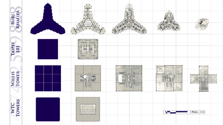
Skyscraper in Dubai, United Arab Emirates Burj Dubai redirects here. Not to be confused with Bur Dubai, a district of Dubai. Burj Khalifaبرج خليفةViewed across The Dubai FountainRecord heightTallest in the world since 2009[I]Preceded byTaipei 101General informationStatusCompletedTypeMixed-useArchitectural styleNeo-futurismLocationDubai, United Arab EmiratesAddress1 Sheikh Mohammed bin Rashid BoulevardNamed forSheikh KhalifaConstruction started6 January 2004; 20 years ago&...


Maurizio Marchei Marchei alla Ternana nella stagione 1977-1978 Nazionalità  Italia Altezza 170 cm Peso 68 kg Calcio Ruolo Ala Termine carriera 1982 CarrieraGiovanili 19??-1973 AtalantaSquadre di club1 1972-1974 Atalanta0 (0)1974-1976 Perugia24 (7)1976-1977 Sambenedettese10 (1)1977-1978 Ternana16 (2)1978-1980 Trento31 (0)1980-1981 Chieti7 (1)1981-1982 Nocera Umbra25 (5) 1 I due numeri indicano le presenze e le reti segnate, per le sole partite di campi...


Artikel ini sebatang kara, artinya tidak ada artikel lain yang memiliki pranala balik ke halaman ini.Bantulah menambah pranala ke artikel ini dari artikel yang berhubungan atau coba peralatan pencari pranala.Tag ini diberikan pada Desember 2023. Yuzuki ItoInformasi pribadiNama lengkap Yuzuki ItoTanggal lahir 7 April 1974 (umur 50)Tempat lahir Prefektur Shizuoka, JepangPosisi bermain GelandangKarier senior*Tahun Tim Tampil (Gol)1993-1997 Shimizu S-Pulse 1998-1999 Kawasaki Frontale 2000-20...

Військово-музичне управління Збройних сил України Тип військове формуванняЗасновано 1992Країна  Україна Емблема управління Військово-музичне управління Збройних сил України — структурний підрозділ Генерального штабу Збройних сил України призначений для планува...


I quattro giganti gassosi del sistema solare, in un fotomontaggio che ne rispetta le dimensioni ma non le distanze; dal basso verso l'alto, Giove, Saturno, Urano e Nettuno. Gigante gassoso (denominato anche pianeta gioviano) è un termine astronomico generico, inventato dallo scrittore di fantascienza James Blish e ormai entrato nell'uso comune[1], per descrivere un grosso pianeta che non sia composto prevalentemente da roccia. I giganti gassosi, in realtà, possono avere un nucleo ro...


Libertador General San Martín Entidad subnacional Otros nombres: Libertador, Ledesma Libertador General San MartínLocalización de Libertador General San Martín en Provincia de JujuyCoordenadas 23°48′00″S 64°47′00″O / -23.8, -64.783333333333Entidad Ciudad y Municipio • País  Argentina • Provincia  Jujuy • Departamento LedesmaIntendente Oscar Jayat (UCR), Partido Frente Cambia JujuyEventos históricos   • Fundación 28 de...

This article needs additional citations for verification. Please help improve this article by adding citations to reliable sources. Unsourced material may be challenged and removed.Find sources: Woodbury Kane – news · newspapers · books · scholar · JSTOR (June 2009) (Learn how and when to remove this message) Woodbury KaneBorn(1859-02-08)February 8, 1859Newport, Rhode Island, U.S.DiedDecember 5, 1905(1905-12-05) (aged 46)New York City, U.S.Educati...


2006 novel by Jabbour Douaihy For the British pharmacologist, see June Raine. June Rain AuthorJabbour DouaihyLanguageArabicGenreCrime fictionSet inNorthern LebanonPublisherDar Al-SaqiPublication date2006Publication placeLebanonPages352 June Rain (Arabic: مطر حزيران)[1] is a 2006 novel written by Lebanese critic and writer Jabbour Douaihy. The novel was published in 2006 by Dar Al-Nahar for Publishing and Distribution by Dar Al-Saqi’s in London. It has been translated t...


Town in Alberta, CanadaStettlerTownTown of StettlerMain Street, StettlerNickname: The Heart of AlbertaStettlerShow map of the County of StettlerStettlerShow map of AlbertaCoordinates: 52°19′25″N 112°43′09″W / 52.32361°N 112.71917°W / 52.32361; -112.71917CountryCanadaProvinceAlbertaRegionCentral AlbertaMunicipal districtCounty of Stettler No. 6Incorporated[1]  • VillageJune 30, 1906 • TownNovember 23, 1906Government...

Lituania memiliki 103 kota. Kota-kota berikut didaftarkan menurut jumlah penduduk Vilnius – 541.278 Kaunas – 364.059 Klaipėda – 188.767 Šiauliai – 130.020 Panevėžys – 116.247 Alytus – 69.859 Marijampolė – 47.693 Mažeikiai – 41.389 Jonava – 34.782 Utena – 33.086 Kėdainiai – 31.613 Telšiai – 30.539 Tauragė – 28.504 Visaginas – 28.438 Ukmergė – 28.006 Plungė – 23.246 Kretinga – 21.425 Šilutė – 21.258 Radviliškis – 19.883 Palanga – 17.611 Drus...


Alice Herdan-Zuckmayer (* 4. April 1901 in Wien, Österreich-Ungarn; † 11. März 1991 in Visp, Schweiz) war eine österreichische Schriftstellerin. Sie war in zweiter Ehe mit Carl Zuckmayer verheiratet. Inhaltsverzeichnis 1 Familiärer Hintergrund 2 Leben 3 Werke 3.1 Briefwechsel 4 Literatur 5 Weblinks 6 Einzelnachweise Familiärer Hintergrund Alice Henriette Alberte Herdan wurde am 4. April 1901 in Wien als Tochter des Juristen Maurice Herdan und der Schauspielerin am Wiener Burgtheater Cl...


Old dump for domestic waste This article is about archaeological remains, known in Spanish as conchales. For the municipality in São Paulo, Brazil, see Conchal. For other uses, see Midden (disambiguation). A closeup of a shell midden in Santa Cruz Province, Argentina. A midden[a] is an old dump for domestic waste.[1] It may consist of animal bones, human excrement, botanical material, mollusc shells, potsherds, lithics (especially debitage), and other artifacts and ecofacts a...

For the book series, see History of Technology (book series). For the academic discipline, see History of science and technology. The wheel, invented sometime before the 4th millennium BC, is one of the most ubiquitous and important technologies. This detail of the Standard of Ur, c. 2500 BCE., displays a Sumerian chariot. History of technology By technological eras Premodern / Pre-industrial Prehistoric Stone Age (lithic) Neolithic Revolution Copper Age Bronze Age Iron Age Ancient Modern Pr...


Pour les articles homonymes, voir Roma. Roma L'entrée de la maison où se déroule une partie de l'intrigue de Roma, à Mexico. Données clés Réalisation Alfonso Cuarón Scénario Alfonso Cuarón Acteurs principaux Yalitza AparicioMarina de Tavira Sociétés de production Esperanto FilmojParticipant Media Pays de production Mexique États-Unis Genre Drame Durée 135 minutes Sortie 2018 Pour plus de détails, voir Fiche technique et Distribution. modifier Roma est un film américano-mexica...